. 2012 Mar;61(1):94-100.
doi: 10.1016/j.parint.2011.07.012. Epub 2011 Jul 19.

Cloning, expression, and characterization of a novel Opisthorchis viverrini calcium-binding EF-hand protein

Affiliations

Cloning, expression, and characterization of a novel Opisthorchis viverrini calcium-binding EF-hand protein

Gulsiri Senawong et al. Parasitol Int. 2012 Mar.

Abstract

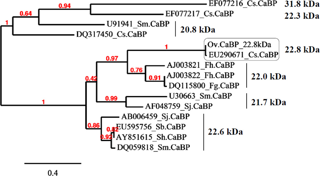
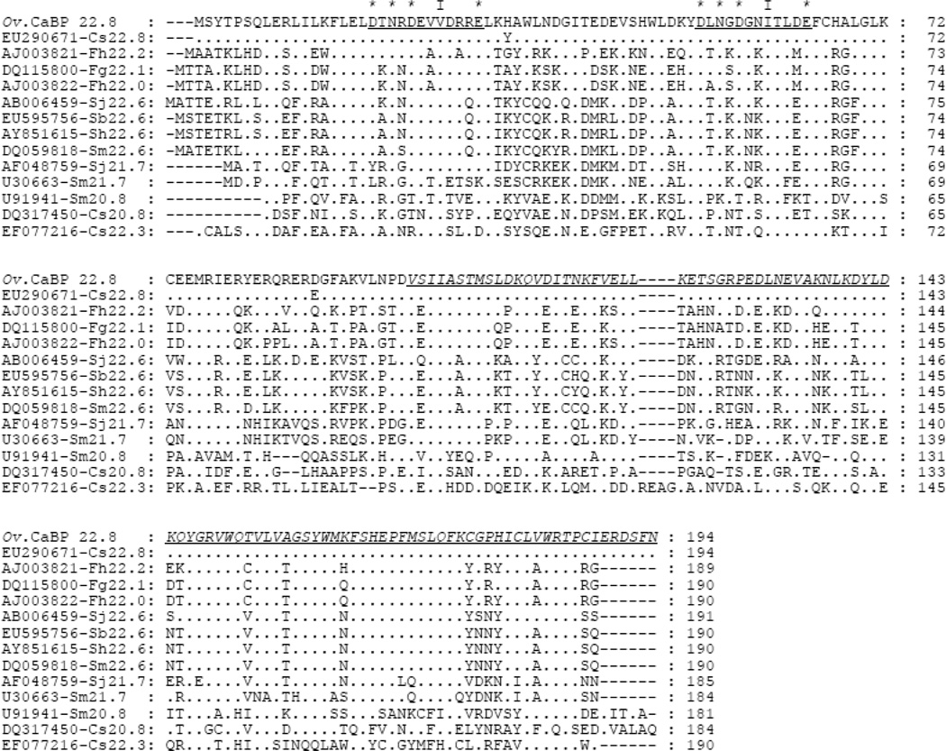
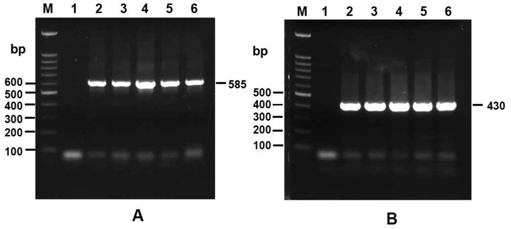
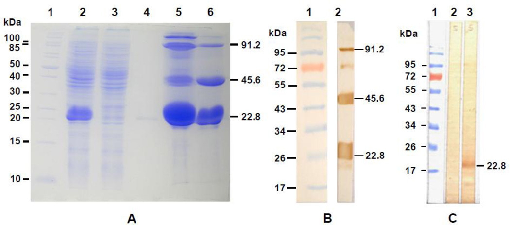
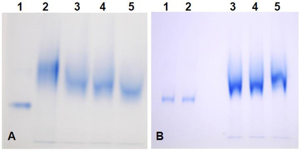
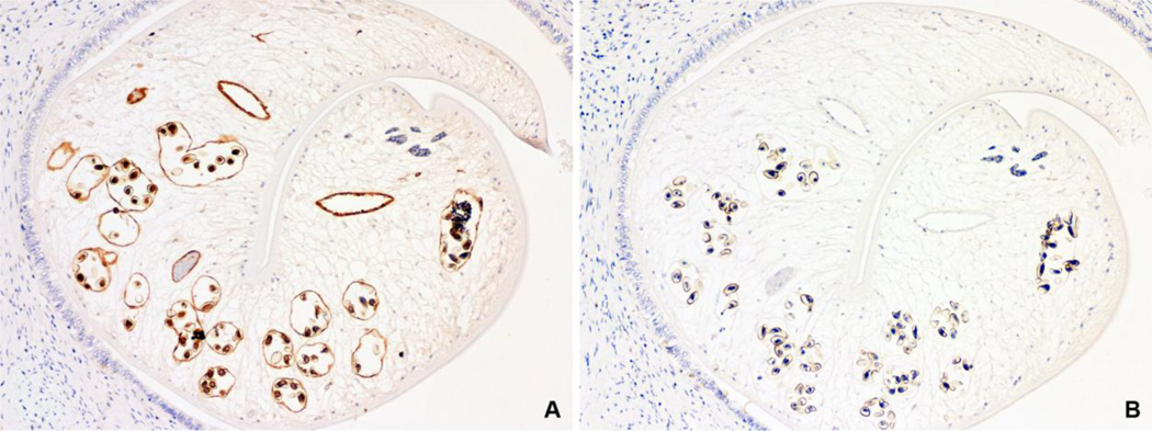
A novel 22.8 kDa of Opisthorchis viverrini (Ov) calcium-binding EF-hand protein (Ov CaBP) was identified and isolated from an immunoscreening of the adult stage Ov cDNA library by using a human cholangiocarcinoma (CCA) serum. This protein was related to other calcium-binding proteins and conserved among the trematodes. Ov CaBP shared 98% amino acid identity to 22.8 kDa of Clonorchis sinensis CaBP and both were classified as a new group of CaBP EF-hand protein by multiple sequence alignment and phylogenetic tree analysis. The open reading frame of Ov CaBP was 585 bp which encoded for 194 amino acids. The N-terminal part is composed of two calcium-binding EF-hand motifs whereas the C-terminal part contains a dynein light chain motif (DLC). In addition, transcription analysis by RT-PCR revealed that it was constitutively transcribed in all stages, including metacercariae, juvenile, and adult. Furthermore, recombinant Ov CaBP protein (rOv CaBP) was expressed as a soluble protein and antibody generated against this rOv CaBP protein was capable of detecting Ov CaBP in the Ov somatic extracts but not in Ov ES products. This anti-rOv CaBP serum was also used to localize Ov CaBP in Ov infected hamster's liver sections which the distribution of Ov CaBP was located in gut epithelium, miracidia in eggs and slightly in parenchyma. Moreover, rOv CaBP protein showed a calcium-binding property in non-denaturing gel mobility shift assay.


Figures

Fig. 1
Fig. 1
The phylogenetic tree of Ov CaBP with homologs from other parasites. GenBank accession number is given followed by abbreviation of species name and the right hand side indicates the molecular weight of CaBPs.
Fig. 2
Fig. 2
Multiple sequence alignment between Ov CaBP and the homologs from Clonorchis, Fasciola, and Schistosoma. GenBank accession numbers are given followed by abbreviation of species name and molecular weight of CaBPs. The underlined sequences indicate the loop region between helix-loop-helix motif of EF-hand 1 and 2. The (*) represents the amino acid residues interacting with calcium while (I) represents the conserved hydrophobic residue within loop whereas the underlined italic letters indicated the DLC part of CaBP.
Fig. 3
Fig. 3
Transcriptional analysis of stage specific expression of Ov CaBP gene by RT-PCR. Lane M, 1, 2, 3, 4, 5, and 6 show a molecular weight marker, negative control (without template), metacercariae, 2-week-old juvenile, 3-week-old juvenile, 1-month-old juvenile, and 2-month-old adult worm, respectively. Panel A shows the RT-PCR amplification of Ov CaBP gene in each stage whereas panel B shows the amplification of actin gene in each stage as an internal control. Electrophoresis was performed on 1% agarose gel.
Fig. 4
Fig. 4
SDS-PAGE of rOv CaBP and western blot analysis of the purified rOv CaBP protein and native Ov CaBP in Ov somatic extracts. In panel A, rOv CaBP protein was purified under the native condition and loaded on 12.5% SDS-PAGE. Lane 1: molecular weight marker, Lane 2: E. coli lysate, Lane 3: flow through, Lane 4–6: eluted fraction 1, 2, and 3 of rOv CaBP protein, respectively. The multimeric forms of rOv CaBP protein were observed and the sizes are shown by arrows. In panel B, western blot was performed to verify the multimer of rOv CaBP protein in the eluted fraction 3 using anti-His tag antibody. Lane 1: molecular weight marker, Lane 2: eluted fraction 3 of purified rOv CaBP protein. In panel C, western blot analysis of native Ov CaBP protein in Ov somatic extracts probed with sera from mice. Lane 1: molecular weight marker, Lane 2: Ov somatic extracts reacted with mouse preimmune serum, Lane 3: Ov somatic extracts reacted with serum from rOv CaBP protein immunized mouse.
Fig. 5
Fig. 5
Analysis of rOv CaBP calcium-binding property by gel mobility shift assay (12.5% PAGE). In panel A, a retardation effect was observed before adding CaCl2 to the protein sample due to the contamination of Ca2+ ion in system, consequently, various concentration of EDTA was added to protein sample for chelating out Ca2+. Lane 1: BSA served as control, Lane 2: rOv CaBP without adding EDTA, Lane 3: rOv CaBP with 1 mM EDTA, Lane 4: rOv CaBP with 5 mM EDTA, Lane 5: rOv CaBP with 20 mM EDTA. In panel B, the mobility shift assay was performed by adding 20 mM EDTA to all protein samples loaded on 12.5% PAGE in the presence/absence of 1 mM MgCl2 or 1 mM CaCl2. Lane 1: BSA with 1 mM MgCl2, Lane 2: BSA with 1 mM CaCl2, Lane 3: rOv CaBP as negative control, Lane 4: rOv CaBP with 1 mM MgCl2, Lane 5: the retardation effect of rOv CaBP with 1 mM CaCl2.
Fig. 6
Fig. 6
Immunolocalization of native Ov CaBP in tissue section of Ov infected hamster’s liver by mouse anti-rOv CaBP serum. Positive immunoperoxidase staining was observed in gut epithelium, miracidia in eggs, and slightly in parenchyma (A), while negative control with mouse preimmune serum showed no staining (B). (Immunoperoxidase, original magnification, ×100)

Similar articles

Cited by

References

    1. Jongsuksuntigul P, Imsomboon T. Opisthorchiasis control in Thailand. Acta Trop. 2003;88:229–232. - PubMed
    1. Sripa B, Kaewkes S, Intapan PM, et al. Food-borne trematodiases in Southeast Asia epidemiology, pathology, clinical manifestation and control. Adv Parasitol. 2010;72:305–350. - PubMed
    1. Bouvard V, Baan R, Straif K, et al. A review of human carcinogens-Part B: biological agents. Lancet Oncol. 2009;10:321–322. - PubMed
    1. Vatanasapt V, Uttaravichien T, Mairiang EO, et al. Cholangiocarcinoma in north-east Thailand. Lancet. 1990;335:116–117. - PubMed
    1. Laha T, Pinlaor P, Mulvenna J, et al. Gene discovery for the carcinogenic human liver fluke, Opisthorchis viverrini. BMC Genomics. 2007;8:189. - PMC - PubMed

Publication types