Comparative Study
. 2011 Sep;150(3):379-89.
doi: 10.1016/j.surg.2011.06.008. Epub 2011 Jul 23.

Efferent vagal nerve stimulation attenuates acute lung injury following burn: The importance of the gut-lung axis

Affiliations
Comparative Study

Efferent vagal nerve stimulation attenuates acute lung injury following burn: The importance of the gut-lung axis

Michael J Krzyzaniak et al. Surgery. 2011 Sep.

Abstract

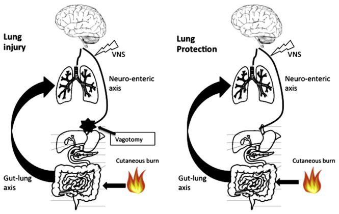
Background: The purpose of this study was to assess acute lung injury when protection to the gut mucosal barrier offered by vagus nerve stimulation is eliminated by an abdominal vagotomy.

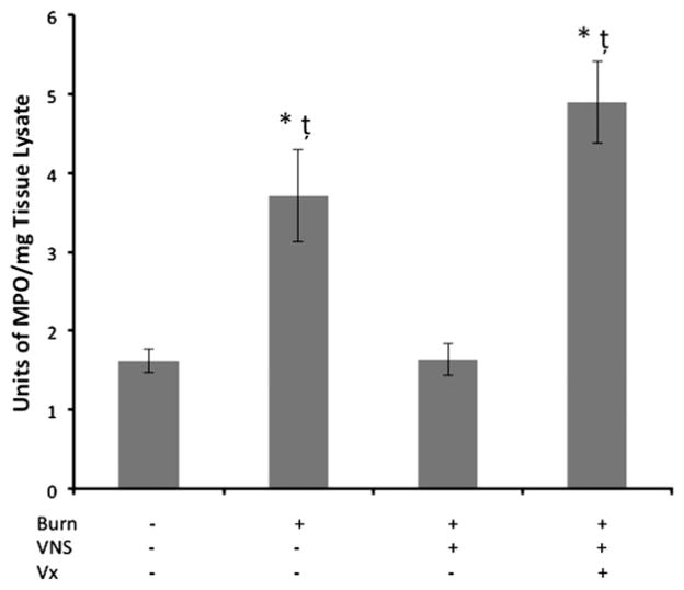
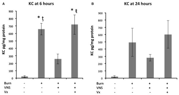
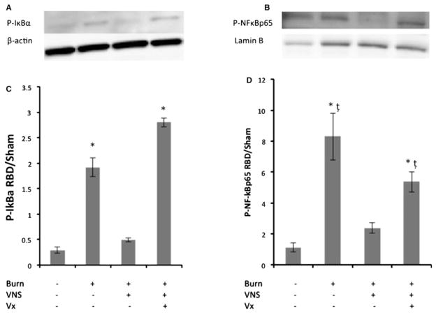
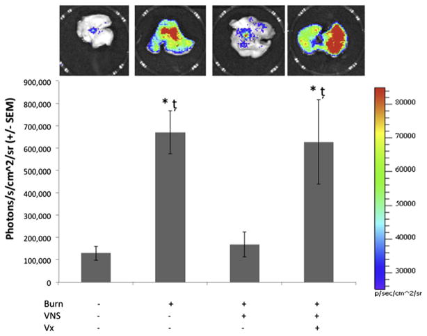
Methods: Male balb/c mice were subjected to 30% total body surface area steam burn with and without electrical stimulation to the right cervical vagus nerve. A cohort of animals were subjected to abdominal vagotomy. Lung histology, myeloperoxidase and ICAM-1 immune staining, myeloperoxidase enzymatic assay, and tissue KC levels were analyzed 24 hours after burn. Additionally, lung IkB-α, NF-kB immunoblots, and NF-kB-DNA binding measured by photon emission analysis using NF-kB-luc transgenic mice were performed.

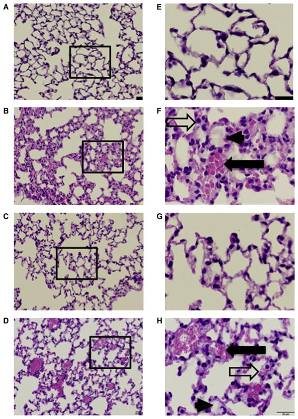
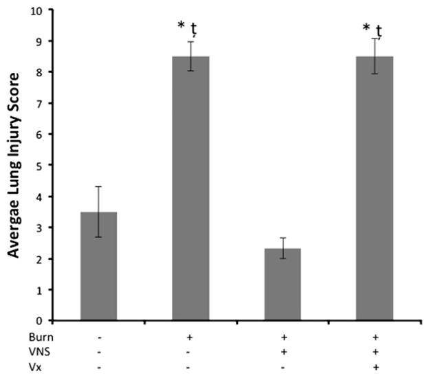
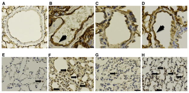
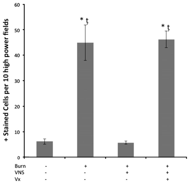
Results: Six hours post burn, phosphorylation of both NF-kB p65 and IkB-α were observed. Increased photon emission signal was seen in the lungs of NF-kB-luc transgenic animals. Vagal nerve stimulation blunted NF-kB activation similar to sham animals whereas abdominal vagotomy eliminated the anti-inflammatory effect. After burn, MPO positive cells and ICAM-1 expression in the lung endothelium was increased, and lung histology demonstrated significant injury at 24 hours. Vagal nerve stimulation markedly decreased neutrophil infiltration as demonstrated by MPO immune staining and enzyme activity. Vagal stimulation also markedly attenuated acute lung injury at 24 hours. The protective effects of vagal nerve stimulation were reversed by performing an abdominal vagotomy.

Conclusion: Vagal nerve stimulation is an effective strategy to protect against acute lung injury following burn. Moreover, the protective effects of vagal nerve stimulation in the prevention of acute lung injury are eliminated by performing an abdominal vagotomy. These results establish the importance of the gut-lung axis after burn in the genesis of acute lung injury.


Figures

Fig. 1
Fig. 1
Lung histology at 24 hours. VNS attenuates burn-induced ALI. Lung sections were harvested from animals after 30% TBSA burn (n ≥ 3 animals per group) then stained with hematoxylin and eosin. Black box denotes the area of the image to the right (E–H). Panels A and E show sections of lung from sham animal at low and high magnification (20× and 60×, respectively). Panels B and F show lung of burned animals showing increased intra-alveolar hemorrhage (solid arrow), thickening of the alveolar membranes (outline arrow) and hyaline membrane formation (arrowhead). Lung sections taken from animals that underwent right cervical VNS prior to injury demonstrate minimal change compared to sham animals as seen in Panels C and G. Panels D and H show sections from animals subjected to abdominal vagotomy prior to VNS and burn. These images demonstrate ALI after the loss of protection from VNS on the gut, indicating that when the gut barrier breaks down, ALI ensues. Black bar: 20 μm.
Fig. 2
Fig. 2
Lung injury score. Lung injury scores were significantly higher in both burn animals and animals subjected to vagotomy prior to VNS and burn. *P < .05 vs sham; t̨P <.05 vs VNS + burn (n ≥ 3 animals per group).
Fig. 3
Fig. 3
ICAM-1and MPO immunostaining at 24 hours after burn. ICAM-1 and MPO expression in lung sections were low in sham and reduced in VNS + burn animals (Panels A, C). and higher following burn (Panel B) and vagotomy + VNS + burn (Panel D). The arrowhead on Panels B and D show increased ICAM-1 deposition on pulmonary endothelium. Neutrophils were identified after staining for MPO and using DAB substrate kit to produce positively stained cells seen with granular appearance (Panels E–H, solid arrows). All images are for ICAM-1 are at 60× magnification and images for MPO are at 40×. Black bar: 20 μm.
Fig. 4
Fig. 4
Neutrophil infiltration in lung tissue at 24 hours. Sham animals had very few neutrophils present in the lungs (6.13 ± 1.1 positively stained cells/10 hpf). Neutrophil infiltration was significantly increased in burn animals (44.9 ± 7 positively stained cells/10 hpf) compared to both sham and VNS + burn animals (5.7 ± 0.6 positively stained cells/10 hpf; *P <.001). Abdominal vagotomy performed prior to VNS + burn causes a significant increase in neutrophil influx similar to burn (46.2 ± 3.2 positively stained cells/10 hpf; t̨P < .001 vs sham and VNS + burn). n ≥ 5 animals per group.
Fig. 5
Fig. 5
MPO activity in lung tissue 24 hours after burn. Myeloperoxidase activity was measured 24 hours after burn and was reduced with VNS prior to burn injury. Sham and VNS + burn groups were similar and were significantly lower than burn alone and when abdominal vagotomy was performed prior to VNS + burn. *P < .005 vs sham; t̨P > .005 vs VNS + burn; n ≥ 5 animals per group.
Fig. 6
Fig. 6
Keratinocyte-derived chemoattractant (KC) levels in lung tissue. KC was measured by ELISA (n ≥ 4 animals per group) 6 and 24 hours after thermal insult. At 6 hours (A) both burn animals and animals subjected to abdominal vagotomy prior to VNS + burn had KC levels significantly higher than sham (*P < .001). VNS + burn animals had levels similar to sham. KC levels were significantly lower in VNS + burn animals than in burn and vagotomized animals (t̨P < .03; t̴P < .01). At 24 hours (B), there was no statistical difference between all groups. Although the trends appear the same, there are modest reductions in KC levels 24 hours after injury. n ≥ 5 animals per group.
Fig. 7
Fig. 7
Cytoplasmic IκB-α and Nuclear NF-κB p65 phosphorylation in lung tissue. Panels A & B show representative Western blots for cytoplasmic P-IκB-α (A) and nuclear NF-κB p65 (B). β-actin and Lamin B loading controls are also shown to demonstrate relatively equal protein load across all lanes. VNS resulted in a decrease in phosphorylation of IκB-α compared to burn alone and animals subjected to vagotomy prior to VNS + burn at 6 hours (*P ≤.001, Panel C). Burn and vagotomized animals also resulted in greater phosphorylation of NF-κB p65 in burn animals compared to sham and VNS + Burn. (*P < .001; t̨P ≤ .01, respectively; Panel D). n ≥ 5 animals per group.
Fig. 8
Fig. 8
Bioluminescence from NF-κB-luc transgenic mice in lung tissue. NF-κB-luc transgenic mice were used to perform luminescent quantification of NF-κB activation in the lungs 6 hours after thermal insult. The images are color matched on the same scale for all animals (n ≥ 5 animals per group). Red signifies more intensity and violet signifies lower intensity. Burn insult produced a near 6-fold increase in luminescence over sham and VNS + burn. Abdominal vagotomy prior to VNS + burn significantly increased luminescent signal compared to sham and VNS + burn. *P ≤ .02 vs sham; t̨P < .03 vs VNS + burn.
Fig. 9
Fig. 9
The importance of the gut-lung axis. This diagram visually represents the importance of the gut-lung axis. When VNS is interrupted by abdominal vagotomy, subsequent ALI occurs as a result of disruption of the neuro-enteric axis leading to intestinal barrier breakdown. If the vagus nerve and the neuro-enteric axis are intact, however, subsequent ALI does not occur in a severe burn model.

References

    1. Pruitt BA, Jr, Erickson DR, Morris A. Progressive pulmonary insufficiency and other pulmonary complications of thermal injury. J Trauma. 1975;15:369–79. - PubMed
    1. Zambon M, Vincent JL. Mortality rates for patients with acute lung injury/ARDS have decreased over time. Chest. 2008;133:1120–7. - PubMed
    1. Bersten AD, Edibam C, Hunt T, Moran J Australian and New Zealand Intensive Care Society Clinical Trials Group. Incidence and mortality of acute lung injury and the acute respiratory distress syndrome in three Australian States. Am J Respir Crit Care Med. 2002;165:443–8. - PubMed
    1. Estenssoro E, Dubin A, Laffaire E, Canales H, Sáenz G, Moseinco M, et al. Incidence, clinical course, and outcome in 217 patients with acute respiratory distress syndrome. Crit Care Med. 2002;30:2450–6. - PubMed
    1. Rubenfeld GD, Caldwell E, Peabody E, Weaver J, Martin DP, Neff M, et al. Incidence and outcomes of acute lung injury. N Engl J Med. 2005;353:1685–93. - PubMed

Publication types

MeSH terms

Substances