. 2011 Sep-Oct;33(5):530-7.
doi: 10.1016/j.ntt.2011.07.003. Epub 2011 Jul 19.

Reduced expression of MAPK/ERK genes in perinatal arsenic-exposed offspring induced by glucocorticoid receptor deficits

Affiliations

Reduced expression of MAPK/ERK genes in perinatal arsenic-exposed offspring induced by glucocorticoid receptor deficits

Ebany J Martinez-Finley et al. Neurotoxicol Teratol. 2011 Sep-Oct.

Abstract

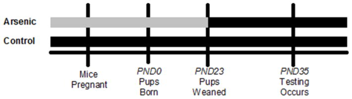
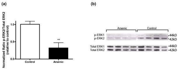
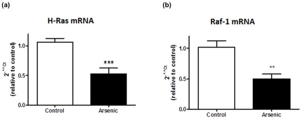
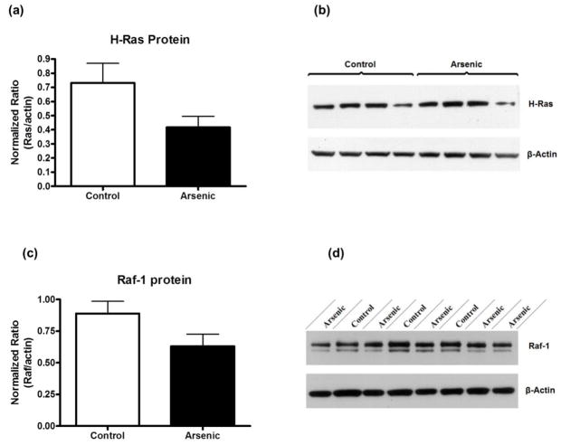
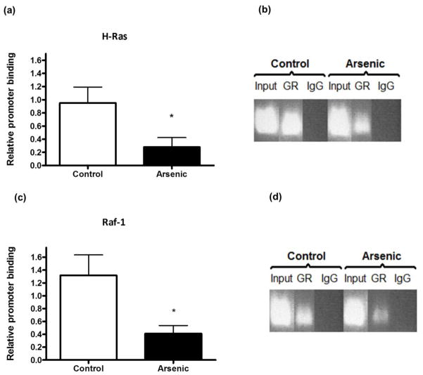
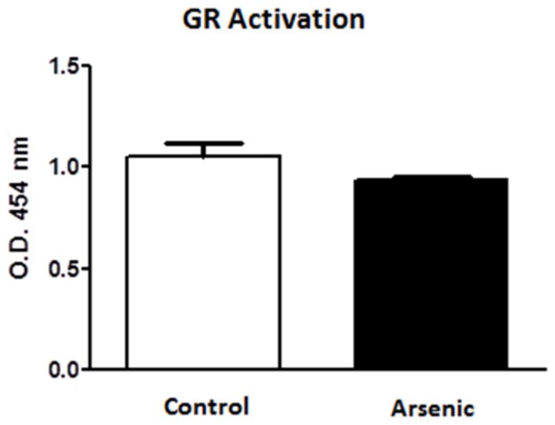
Changes within the glucocorticoid receptor (GR) cellular signaling pathway were evaluated in adolescent mice exposed to 50 ppb arsenic during gestation. Previously, we reported increased basal plasma corticosterone levels, decreased hippocampal GR levels and deficits in learning and memory performance in perinatal arsenic-exposed mice. The biosynthesis of members of the mitogen-activated protein kinase (MAPK) signaling pathway, known to be involved in learning and memory, is modulated by the binding of GR to glucocorticoid response elements (GREs) in the gene promoters. Two genes of the MAPK pathway, Ras and Raf, contain GREs which are activated upon binding of GRs. We evaluated the activity of GRs at Ras and Raf promoters using chromatin immunoprecipitation and real-time PCR and report decreased binding of the GR at these promoters. An ELISA-based GR binding assay was used to explore whether this decreased binding was restricted to in vivo promoters and revealed no differences in binding of native GR to synthetic GREs. The decreased in vivo GR binding coincides with significantly decreased mRNA levels and slight reductions of protein of both H-Ras and Raf-1 in perinatally arsenic-exposed mice. Nuclear activated extracellular-signal regulated kinase (ERK), a downstream target of Ras and Raf, whose transcriptional targets also play an important role in learning and memory, was decreased in the hippocampus of arsenic-exposed animals when compared to controls. GR-mediated transcriptional deficits in the MAPK/ERK pathway could be an underlying cause of previously reported learning deficits and provide the link to arsenic-induced deficiencies in cognitive development.


Figures

Figure 1
Figure 1
Perinatal Arsenic Exposure Paradigm. Black bars represent standard tap water [5 ppb]. Grey bars represent arsenic dissolved in standard tap water [55ppb]. Female mice were started on the arsenic or control water 2 weeks prior to pregnancy. Arsenic exposed pups were weaned at PND23 and transferred to standard tap water until experimentation at PND35. PND=postnatal day
Figure 2
Figure 2
Activated ERK in hippocampal nuclear subcellular fraction. (a) Quantitation of p-ERK, total ERK, and β-actin bands in hippocampal nuclear subcellular fraction of arsenic exposed mice was based on densitometric analysis (**p = 0.003 vs. control). Results are expressed as a percentage of the p-ERK2 signal to total ERK2 signal, relative to control animals, and are presented as mean ± SEM of five-six litters. (b) Representative western blot of p-ERK1/2 and total ERK1/2 in the nuclear compartment of perinatal exposed and control mice.
Figure 3
Figure 3
Reduced MAPK pathway mRNA levels in perinatal arsenic-exposed offspring compared to controls. (a) H-Ras mRNA levels in arsenic-exposed offspring and controls (***p = 0.0005 vs. control). (b) Raf-1 mRNA levels in arsenic-exposed offspring and controls (**p = 0.003 vs. control). Results are expressed as a percentage of the H-Ras or Raf-1 signal to the β-actin signal, relative to control animals, and are presented as mean ± SEM of eight-ten litters.
Figure 4
Figure 4
MAPK pathway protein levels were only slightly reduced in perinatal arsenic-exposed offspring compared to controls. (a) H-Ras protein levels in arsenic-exposed offspring and controls (p = 0.098 vs. control). (b) Representative western blot of H-Ras and β-actin. (c) Raf-1 protein levels in arsenic-exposed offspring and controls (p = 0.07 vs. control). (d) Representative western blot of Raf-1 and β-actin. Results are expressed as a percentage of the H-Ras or Raf-1 signal to the β-actin signal, relative to control animals, and are presented as mean ± SEM of ten litters.
Figure 5
Figure 5
Perinatal arsenic exposure reduces glucocorticoid receptor (GR) binding to H-Ras and Raf-1 promoters. (a) Results of chromatin immunoprecipitation with anti-GR antibody in nuclear tissue extracts followed by real-time PCR for H-Ras (*p = 0.04 vs. control). (b) Gel showing real-time PCR product bands for Input (positive control), GR and IgG (negative control). (c) Results of chromatin immunoprecipitation with anti-GR antibody in nuclear tissue extracts followed by real-time PCR for Raf-1 (p = 0.06 vs. control). (d) Gel showing real-time PCR bands for Input (positive control), GR and IgG (negative control). Results are presented as relative promoter binding, relative to controls, after subtracting for non-immune rabbit IgG interactions. Results are representative of five separate experiments and are presented as mean ± SEM of four-five litters. GR: Glucocorticoid Receptor; IgG: normal rabbit IgG
Figure 6
Figure 6
Perinatal arsenic does not interfere with the binding of native GR to a consensus GRE sequence. Assay was conducted three separate times. Data are mean optical density units (454 nm) ± SEM of eight litters.

References

    1. Agency for Toxic Substances and Disease Registry (ATSDR) Toxicological profile for Arsenic. Atlanta, GA: U.S. Department of Health and Human Services, Public Health Service; 2007. [Accessed 05 Jan 2008]. http://www.atsdr.cdc.gov/toxprofiles/tp2.html.
    1. Atkins CM, Selcher JC, Petraitis JJ, Trzaskos JM, Sweatt JD. The MAPK cascade is required for mammalian associative learning. Nat Neurosci. 1998;1:602–9. - PubMed
    1. Beato M, Chalepakis G, Schauer M, Slater EP. DNA regulatory elements for steroid hormones. J Steroid Biochem. 1989;32:737–47. - PubMed
    1. Blum S, Moore AN, Adams F, Dash PK. A mitogen-activated protein kinase cascade in the CA1/CA2 subfield of the dorsal hippocampus is essential for long-term spatial memory. J Neurosci. 1999;19:3535–44. - PMC - PubMed
    1. Bodwell JE, Gosse JA, Nomikos AP, Hamilton JW. Arsenic Disruption of Steroid Receptor Gene Activation: Complex Dose-Response Effects Are Shared by Several Steroid Receptors. Chem Res Toxicol. 2006;19:1619–29. - PMC - PubMed

Publication types

MeSH terms

Substances