

Biomaterial implants mediate autologous stem cell recruitment in mice
- PMID: 21784181
- PMCID: PMC3185118
- DOI: 10.1016/j.actbio.2011.06.050
Biomaterial implants mediate autologous stem cell recruitment in mice
Abstract
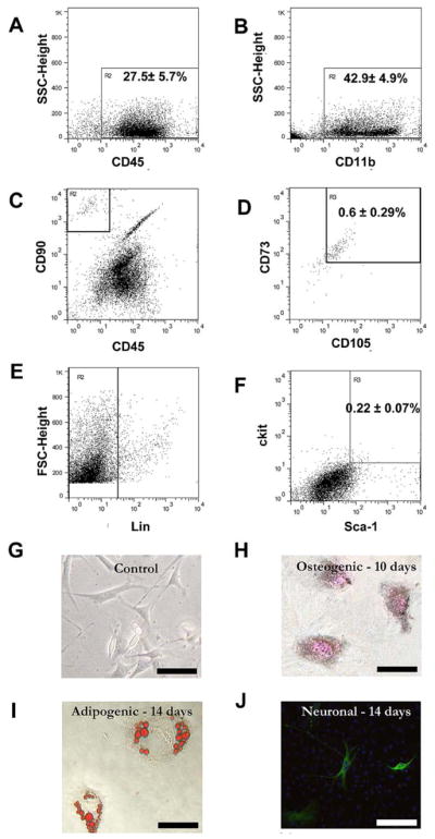
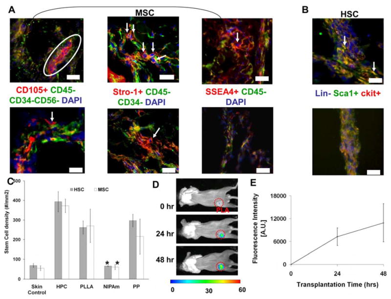
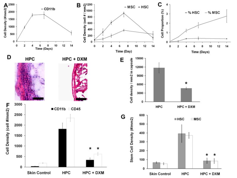
Autologous stem cells, recognized as the best cells for stem cell therapy, are associated with difficult extraction procedures which often lead to more traumas for the patients and time-consuming laboratory work, which delays their subsequent application. To combat such challenges, it was recently uncovered that, shortly after biomaterial implantation, following the recruitment of inflammatory cells, substantial numbers of mesenchymal stem cells (MSC) and hematopoietic stem cells (HSC) were recruited to the implantation sites. These multipotent MSC could be differentiated into various lineages in vitro. Inflammatory signals may be responsible for the gathering of stem cells, since there is a good relationship between biomaterial-mediated inflammatory responses and stem cell accumulation in vivo. In addition, the treatment with the anti-inflammatory drug dexamethasone substantially reduced the recruitment of both MSC and HSC. The results from this work support that such strategies could be further developed towards localized recruitment and differentiation of progenitor cells. This may permit the future development of autologous stem cell therapies without the need for tedious cell isolation, culture and transplantation.
Copyright © 2011 Acta Materialia Inc. Published by Elsevier Ltd. All rights reserved.
Figures
Similar articles
-
The effect of incorporation of SDF-1alpha into PLGA scaffolds on stem cell recruitment and the inflammatory response.Biomaterials. 2010 May;31(14):3997-4008. doi: 10.1016/j.biomaterials.2010.01.144. Epub 2010 Feb 24. Biomaterials. 2010. PMID: 20185171 Free PMC article.
-
Current approaches in biomaterial-based hematopoietic stem cell niches.Acta Biomater. 2018 May;72:1-15. doi: 10.1016/j.actbio.2018.03.028. Epub 2018 Mar 22. Acta Biomater. 2018. PMID: 29578087 Review.
-
Mobilization of hematopoietic stem cells for use in autologous transplantation.Clin J Oncol Nurs. 2010 Apr;14(2):212-22. doi: 10.1188/10.CJON.212-222. Clin J Oncol Nurs. 2010. PMID: 20350895 Review.
-
Mesenchymal stromal cells induce a permissive state in the bone marrow that enhances G-CSF-induced hematopoietic stem cell mobilization in mice.Exp Hematol. 2018 Aug;64:59-70.e2. doi: 10.1016/j.exphem.2018.05.002. Exp Hematol. 2018. PMID: 29775645
-
Etoposide-mediated interleukin-8 secretion from bone marrow stromal cells induces hematopoietic stem cell mobilization.BMC Cancer. 2020 Jul 2;20(1):619. doi: 10.1186/s12885-020-07102-x. BMC Cancer. 2020. PMID: 32615949 Free PMC article.
Cited by
-
Development of optical probes for in vivo imaging of polarized macrophages during foreign body reactions.Acta Biomater. 2014 Jul;10(7):2945-2955. doi: 10.1016/j.actbio.2014.04.001. Epub 2014 Apr 13. Acta Biomater. 2014. PMID: 24726956 Free PMC article.
-
Novel source of human hematopoietic stem cells from peritoneal dialysis effluents.Stem Cell Res. 2015 Sep;15(2):299-304. doi: 10.1016/j.scr.2015.07.003. Epub 2015 Jul 16. Stem Cell Res. 2015. PMID: 26209817 Free PMC article.
-
Preparation of a novel injectable in situ-gelling nanoparticle with applications in controlled protein release and cancer cell entrapment.RSC Adv. 2018 Oct 9;8(60):34625-34633. doi: 10.1039/c8ra06589f. eCollection 2018 Oct 4. RSC Adv. 2018. PMID: 35548629 Free PMC article.
-
Chemokine releasing particle implants for trapping circulating prostate cancer cells.Sci Rep. 2020 Mar 10;10(1):4433. doi: 10.1038/s41598-020-60696-x. Sci Rep. 2020. PMID: 32157115 Free PMC article.
-
Development of a dual-wavelength fluorescent nanoprobe for in vivo and in vitro cell tracking consecutively.Bioorg Med Chem. 2019 May 1;27(9):1855-1862. doi: 10.1016/j.bmc.2019.03.036. Epub 2019 Mar 19. Bioorg Med Chem. 2019. PMID: 30910476 Free PMC article.
References
-
- Bruder SP, Kurth AA, Shea M, Hayes WC, Jaiswal N, Kadiyala S. Bone regeneration by implantation of purified, culture-expanded human mesenchymal stem cells. J Orthop Res. 1998;16:155–62. - PubMed
-
- Loebinger MR, Aguilar S, Janes SM. Therapeutic potential of stem cells in lung disease: progress and pitfalls. Clin Sci (Lond) 2008;114:99–108. - PubMed
-
- Gang EJ, Bosnakovski D, Figueiredo CA, Visser JW, Perlingeiro RC. SSEA-4 identifies mesenchymal stem cells from bone marrow. Blood. 2007;109:1743–51. - PubMed
-
- Li TS, Hayashi M, Ito H, Furutani A, Murata T, Matsuzaki M, Hamano K. Regeneration of infarcted myocardium by intramyocardial implantation of ex vivo transforming growth factor-beta-preprogrammed bone marrow stem cells. Circulation. 2005;111:2438–45. - PubMed
-
- Price MJ, Chou CC, Frantzen M, Miyamoto T, Kar S, Lee S, Shah PK, Martin BJ, Lill M, Forrester JS, et al. Intravenous mesenchymal stem cell therapy early after reperfused acute myocardial infarction improves left ventricular function and alters electrophysiologic properties. Int J Cardiol. 2006;111:231–9. - PubMed
Publication types
MeSH terms
Substances
Grants and funding
LinkOut - more resources
Full Text Sources
Other Literature Sources
Medical
