. 2011 Aug 23:1408:81-7.
doi: 10.1016/j.brainres.2011.06.051. Epub 2011 Jun 26.

Functional significance of aldehyde dehydrogenase ALDH1A1 to the nigrostriatal dopamine system

Affiliations

Functional significance of aldehyde dehydrogenase ALDH1A1 to the nigrostriatal dopamine system

David W Anderson et al. Brain Res. .

Abstract

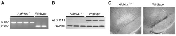
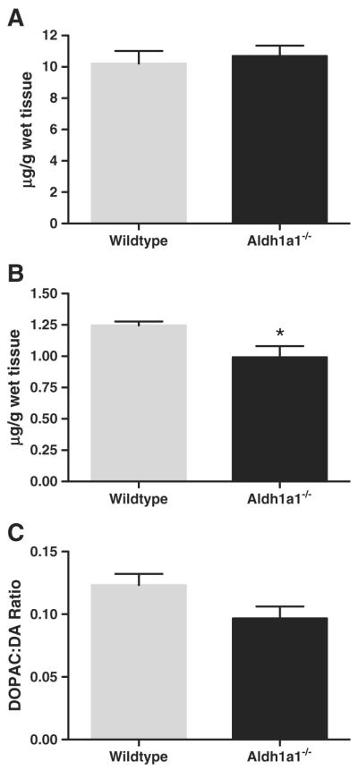
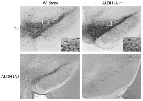
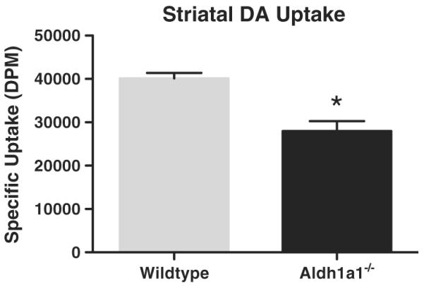
Aldehyde dehydrogenase 1A1 (ALDH1A1) is a member of a superfamily of detoxification enzymes found in various tissues that participate in the oxidation of both aliphatic and aromatic aldehydes. In the brain, ALDH1A1 participates in the metabolism of catecholamines including dopamine (DA) and norepinephrine, but is uniquely expressed in a subset of dopaminergic (DAergic) neurons in the ventral mesencephalon where it converts 3,4-dihydroxyphenylacetaldehyde, a potentially toxic aldehyde, to 3,4-dihydroxyphenylacetic acid, a non toxic metabolite. Therefore, loss of ALDH1A1 expression could be predicted to alter DA metabolism and potentially increase neurotoxicity in ventral mesencephalic DA neurons. Recent reports of reduced levels of expression of both Aldh1a1 mRNA and protein in the substantia nigra (SN) of Parkinson's disease patients suggest possible involvement of ALDH1A1 in this progressive neurodegenerative disease. The present study used an Aldh1a1 null mouse to assess the influence of ALDH1A1 on the function and maintenance of the DAergic system. Results indicate that the absence of Aldh1a1 did not negatively affect growth and development of SN DA neurons nor alter protein expression levels of tyrosine hydroxylase, the DA transporter or vesicular monoamine transporter 2. However, absence of Aldh1a1 significantly increased basal extracellular DA levels, decreased KCl and amphetamine stimulated DA release and decreased DA re-uptake and resulted in more tyrosine hydroxylase expressing neurons in the SN than in wildtype animals. These data suggest that in young adult animals with deletion of the Aldh1a1 gene there is altered DA metabolism and dysfunction of the DA transporter and DA release mechanisms.


Figures

Fig. 1
Fig. 1
Expression of Aldh1a1 mRNA and protein in Aldh1a1−/− and wildtype (WT) mice. (A) Representative genotyping gel. Aldh1a1−/− animals did not contain a functional copy of the Aldh1a1 gene, but were positive for the Neo insert used to disrupt gene expression (NEO cassette, 600 bp; Aldh1a1, 250 bp). (B) Western blot analysis of ALDH1A1 protein in the striatum showed a significant loss of ALDH1A1 expression in the Aldh1a1−/− mice compared to normal expression levels in WT mice. Loading controls (using GAPDH) for all lanes are shown. (C) Immunohistochemical analysis of ALDH1A1 expression in the substantia nigra pars compacta showed significant loss ALDH1A1-immunoreactivity when compared to WT animals.
Fig. 2
Fig. 2
Analysis of dopamine (DA) levels in the striatum of freely moving awake mice by in vivo microdialysis. (A) Basal extracellular levels of DA were significantly higher in the Aldh1a1−/− mice than wildtype (WT) mice. (B) KCl-stimulated DA release was significantly reduced in Aldh1a1−/− mice compared to WT mice. (C) Amphetamine stimulated DA release was significantly reduced in Aldh1a1−/− mice compared to WT mice. *p<0.05 vs. wildtype. (n=8 and 6 respectively for Aldh1a1−/− and WT).
Fig. 3
Fig. 3
Comparison of tissue dopamine (DA) and metabolite levels in the striatum of Aldh1a1−/− and wildtype mice. (A) Striatal DA levels in Aldh1a1−/− and wildtype (WT) mice were not significantly different. (B) The level of 3,4-dihydroxyphenylacetic acid (DOPAC) in the Aldh1a1−/− mouse was significantly lower than in WT mice (*p<0.05). (C) Dopamine turnover was reduced in Aldh1a1−/− mice when compared to WT, but the difference was not statistically significant. (n=10 and 6 respectively for Aldh1a1−/− and WT).
Fig. 4
Fig. 4
Photomicrographs of ALDH1A1 and tyrosine hydroxylase immunoreactivity in the substantia nigra of Aldh1a1−/− and wildtype (WT) mice. All images were taken at 4× magnification, with higher 40× magnification inset for both tyrosine hydroxylase photomicrographs. (n=6 and 6 respectively for Aldh1a1−/− and WT).
Fig. 5
Fig. 5
Striatal protein expression in Aldh1a1−/− and wildtype (WT) mice. There were no significant differences in any of the analyzed proteins between the Aldh1a1−/− and WT mice. TH=tyrosine hydroxylase; DAT=dopamine transporter; VMAT2=vesicular monoamine transporter 2. GAPDH or β-actin was used as loading controls. (n=6 and 6 respectively for Aldh1a1−/− and WT).
Fig. 6
Fig. 6
Analysis of striatal dopamine uptake in Aldh1a1−/− and wildtype (WT) mice. There was a significant decrease in DA uptake in striatal homogenates from Aldh1a1−/− animals compared to wildtype control animals. *p<0.001. (n=7 and 7 respectively for Aldh1a1−/− and WT).

References

    1. Adams JD, Jr., Chang ML, Klaidman L. Parkinson's disease—redox mechanisms. Curr. Med. Chem. 2001;8:809–814. - PubMed
    1. Anderson DW, Neavin T, Smith JA, Schneider JS. Neuroprotective effects of pramipexole in young and aged MPTP-treated mice. Brain Res. 2001;905:44–53. - PubMed
    1. Burke WJ, Li SW, Williams EA, Nonneman R, Zahm DS. 3,4-Dihydroxyphenylacetaldehyde is the toxic dopamine metabolite in vivo: implications for Parkinson's disease pathogenesis. Brain Res. 2003;989:205–213. - PubMed
    1. Erwin VG, Deitrich RA. Brain aldehyde dehydrogenase. Localization, purification, and properties. J. Biol. Chem. 1966;241:3533–3539. - PubMed
    1. Fan X, Molotkov A, Manabe S-I, Donmoyer CM, Deltour L, Foglio MH, Cuenca AE, Blaner WS, Lipton SA, Duester G. Targeted disruption of Aldh1a1 (Raldh1) provides evidence for a complex mechanism of retinoic acid synthesis in the developing retina. Mol. Cell. Biol. 2003;23:4637–4648. - PMC - PubMed

Publication types