

Staphylococcal biofilm disassembly
- PMID: 21784640
- PMCID: PMC3164736
- DOI: 10.1016/j.tim.2011.06.004
Staphylococcal biofilm disassembly
Abstract
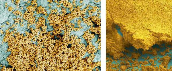
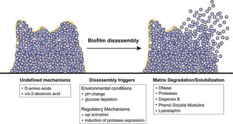
Staphylococcus aureus and Staphylococcus epidermidis are a frequent cause of biofilm-associated infections that are a tremendous burden on our healthcare system. Staphylococcal biofilms exhibit extraordinary resistance to antimicrobial killing, limiting the efficacy of antibiotic therapy, and surgical intervention is often required to remove infected tissues or implanted devices. Recent work has provided new insight into the molecular basis of biofilm development in these opportunistic pathogens. Extracellular bacterial products, environmental conditions, and polymicrobial interactions have all been shown to influence profoundly the ability of these bacteria to colonize and disperse from clinically relevant surfaces. We review new developments in staphylococcal biofilm disassembly and set them in the context of potential strategies to control biofilm infections.
Copyright © 2011 Elsevier Ltd. All rights reserved.
Figures
References
-
- O'Gara JP. ica and beyond: biofilm mechanisms and regulation in Staphylococcus epidermidis and Staphylococcus aureus. FEMS Microbiol. Lett. 2007;270:179–188. - PubMed
-
- Gotz F. Staphylococcus and biofilms. Molecular Microbiology. 2002;43:1367–1378. - PubMed
-
- Parsek MR, Singh PK. Bacterial biofilms: an emerging link to disease pathogenesis. Annu. Rev. Microbiol. 2003;57:677–701. - PubMed
-
- del Pozo JL, Patel R. The challenge of treating biofilm-associated bacterial infections. Clin. Pharmacol. Ther. 2007;82:204–209. - PubMed
Publication types
MeSH terms
Grants and funding
LinkOut - more resources
Full Text Sources
Other Literature Sources
Medical
