. 2011 Sep 16;286(37):32790-800.
doi: 10.1074/jbc.M111.245985. Epub 2011 Jul 22.

Adiponectin ameliorates doxorubicin-induced cardiotoxicity through Akt protein-dependent mechanism

Affiliations

Adiponectin ameliorates doxorubicin-induced cardiotoxicity through Akt protein-dependent mechanism

Sonomi Maruyama et al. J Biol Chem. .

Abstract

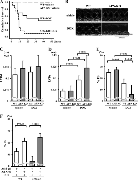
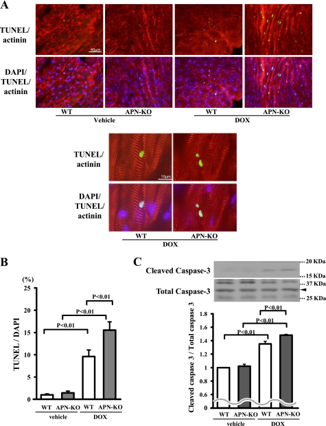
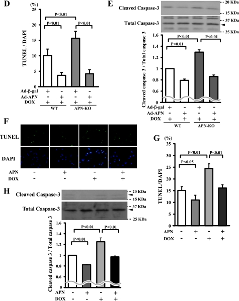
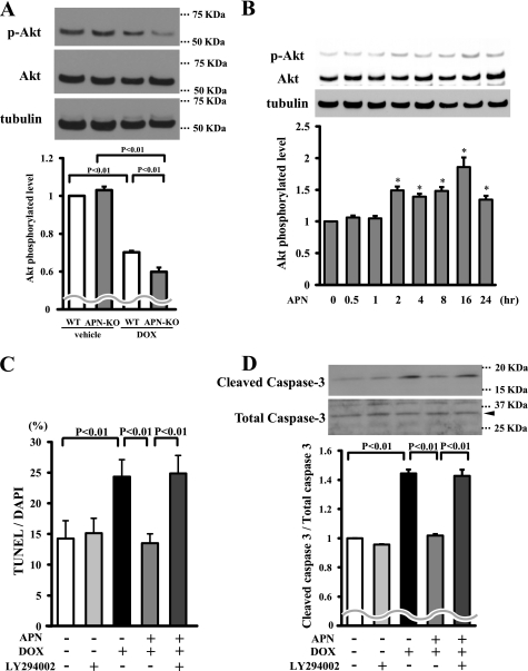
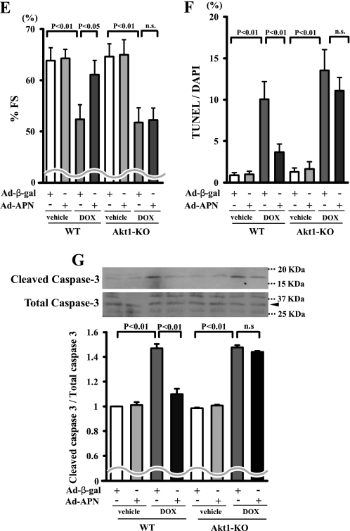
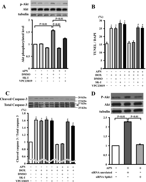
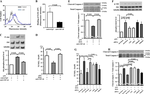
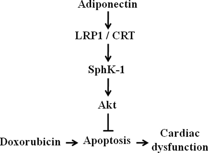
Accumulating evidence shows that obesity is associated with doxorubicin cardiac toxicity in the heart, but the molecular mechanisms that contribute to this pathological response are not understood. Adiponectin is an adipose-derived, cardioprotective factor that is down-regulated in obesity. Here, we investigated the effect of adiponectin on doxorubicin (DOX)-induced cardiotoxicity and assessed the mechanisms of this effect. A single dose of DOX was intraperitoneally injected into the abdomen of adiponectin knock-out (APN-KO) and wild-type (WT) mice. APN-KO mice had increased mortality and exacerbated contractile dysfunction of left ventricle compared with WT mice. APN-KO mice also showed increased apoptotic activity and diminished Akt signaling in the failing myocardium. Systemic delivery of adenoviral vector expressing adiponectin improved left ventricle dysfunction and myocardial apoptosis following DOX injection in WT and APN-KO mice but not in Akt1 heterozygous KO mice. In cultured rat neonatal cardiomyocytes, adiponectin stimulated Akt phosphorylation and inhibited DOX-stimulated apoptosis. Treatment with sphingosine kinase-1 inhibitor or sphingosine 1-phosphate receptor antagonist diminished adiponectin-induced Akt phosphorylation and reversed the inhibitory effects of adiponectin on myocyte apoptosis. Pretreatment with anti-calreticulin antibody reduced the binding of adiponectin to cardiac myocytes and blocked the adiponectin-stimulated increase in Akt activation and survival in cardiomyocytes. Interference of the LRP1/calreticulin co-receptor system by siRNA or blocking antibodies diminished the stimulatory actions of adiponectin on Akt activation and myocyte survival. These data show that adiponectin protects against DOX-induced cardiotoxicity by its ability to promote Akt signaling.


Figures

FIGURE 1.
FIGURE 1.
Adiponectin improves DOX-induced cardiac dysfunction. A, Kaplan-Meier survival analysis of WT and KO mice following DOX or vehicle administration (n = 11 in each group). B, representative M-mode echocardiograms for WT or KO mice at 5 days after DOX or vehicle injection. Vertical scale bar, 1 mm; horizontal scale bar, 200 ms. C–E, quantitative analysis of the LVDd (C), LVDs (D), and %FS (E) for WT and KO mice at 5 days after DOX injection (n = 5). F, quantitative analysis of %FS in WT and APN-KO mice treated with Ad-β-gal or Ad-APN at 5 days after DOX injection. Ad-APN or Ad-β-gal (2 × 10⁸ pfu total) was delivered intravenously via the tail vein 3 days before DOX injection (n = 5). Results are presented as mean ± S.D.
FIGURE 2.
FIGURE 2.
Adiponectin suppresses DOX-induced apoptosis in heart. A, representative photographs of heart sections stained with TUNEL from WT and APN-KO mice at 5 days after DOX or vehicle injection. Apoptotic nuclei were determined by TUNEL staining (green). Myocytes were determined by sarcomeric actinin (red), and total nuclei were counterstained with DAPI (blue). High magnification representative photographs of heart sections are shown in the bottom panels. Scales bars, 40 μm (top) and 10 μm (bottom). B, quantitative analysis of apoptotic nuclei from WT (n = 5) and APN-KO mouse (n = 5) hearts following DOX or vehicle injection. TUNEL-positive nuclei were counted in several randomly selected fields and expressed as a percentage of the total number of nuclei. C, detection of cleaved caspase-3 from WT (n = 5) and APN-KO mouse (n = 5) hearts following DOX or vehicle injection by Western blot analysis. D, quantitative analysis of apoptotic nuclei from WT (n = 5) and APN-KO (n = 5) mouse hearts treated with Ad-APN or Ad-β-gal following DOX injection. TUNEL-positive nuclei were counted in several randomly selected fields and expressed as a percentage of the total number of nuclei. E, detection of cleaved caspase-3 from WT (n = 5) and APN-KO (n = 5) mouse hearts treated with Ad-APN or Ad-β-gal following DOX injection by Western blot analysis. F, effect of DOX on cultured myocytes. Representative photographs of TUNEL-positive cardiac myocytes under conditions of serum deprivation with or without DOX stimulation for 24 h in the presence or absence of adiponectin protein are shown. Apoptotic nuclei were identified by TUNEL staining (green), and total nuclei were identified by DAPI counterstaining (blue). G, quantitative analysis of TUNEL-positive cells with DOX or vehicle exposure in the presence or absence of adiponectin. TUNEL-positive nuclei were counted in several randomly selected fields and expressed as a percentage of the total number of nuclei (n = 4). H, detection of cleaved caspase-3 with DOX or vehicle exposure in the presence or absence of adiponectin.
FIGURE 2.
FIGURE 2.
Adiponectin suppresses DOX-induced apoptosis in heart. A, representative photographs of heart sections stained with TUNEL from WT and APN-KO mice at 5 days after DOX or vehicle injection. Apoptotic nuclei were determined by TUNEL staining (green). Myocytes were determined by sarcomeric actinin (red), and total nuclei were counterstained with DAPI (blue). High magnification representative photographs of heart sections are shown in the bottom panels. Scales bars, 40 μm (top) and 10 μm (bottom). B, quantitative analysis of apoptotic nuclei from WT (n = 5) and APN-KO mouse (n = 5) hearts following DOX or vehicle injection. TUNEL-positive nuclei were counted in several randomly selected fields and expressed as a percentage of the total number of nuclei. C, detection of cleaved caspase-3 from WT (n = 5) and APN-KO mouse (n = 5) hearts following DOX or vehicle injection by Western blot analysis. D, quantitative analysis of apoptotic nuclei from WT (n = 5) and APN-KO (n = 5) mouse hearts treated with Ad-APN or Ad-β-gal following DOX injection. TUNEL-positive nuclei were counted in several randomly selected fields and expressed as a percentage of the total number of nuclei. E, detection of cleaved caspase-3 from WT (n = 5) and APN-KO (n = 5) mouse hearts treated with Ad-APN or Ad-β-gal following DOX injection by Western blot analysis. F, effect of DOX on cultured myocytes. Representative photographs of TUNEL-positive cardiac myocytes under conditions of serum deprivation with or without DOX stimulation for 24 h in the presence or absence of adiponectin protein are shown. Apoptotic nuclei were identified by TUNEL staining (green), and total nuclei were identified by DAPI counterstaining (blue). G, quantitative analysis of TUNEL-positive cells with DOX or vehicle exposure in the presence or absence of adiponectin. TUNEL-positive nuclei were counted in several randomly selected fields and expressed as a percentage of the total number of nuclei (n = 4). H, detection of cleaved caspase-3 with DOX or vehicle exposure in the presence or absence of adiponectin.
FIGURE 3.
FIGURE 3.
Adiponectin inhibits DOX-induced myocyte apoptosis via Akt signaling. A, the representative immunoblots in phosphorylated Akt (p-Akt) in heart tissues from WT and APN-KO mice at 5 days after DOX or vehicle injection (left). Akt phosphorylation levels in myocardium were analyzed by Western blotting. Right, quantitative analysis of relative changes in phosphorylated Akt. Phosphorylation levels of Akt were normalized to the α-tubulin signal and expressed as the percentage of the signal intensity of vehicle-injected WT mice (n = 5). B, time-dependent changes in the phosphorylation of Akt in rat cultured cardiac myocytes after adiponectin treatment (10 μg/ml). Quantitative analysis of relative changes in phosphorylated Akt (n = 4) (*, p < 0.05 versus control) is shown. C and D, involvement of Akt in adiponectin inhibition of DOX-stimulated myocyte apoptosis. Cells were pretreated with the phosphatidylinositol 3-kinase inhibitor LY294002 or vehicle for 60 min and treated with or without adiponectin (10 μg/ml) followed by stimulation with DOX for 24 h. Quantitative analysis of TUNEL-positive nuclei (C) and caspase-3 activity (D) is shown. TUNEL-positive nuclei were counted in several randomly selected fields and expressed as a percentage of the total number of nuclei (n = 4). E, F, and G, effects of adiponectin on DOX-induced cardiomyopathy in Akt-1 KO mice. Quantitative analysis of %FS (E), TUNEL-positive nuclei in hearts (F), and caspase-3 activity (G) in Ad-β-gal- or Ad-APN-treated WT and Akt-1 KO mice at 5 days after DOX injection (n = 5) is shown. Ad-APN or Ad-β-gal (2 × 10⁸ pfu total) was delivered intravenously via the tail vein 3 days before DOX injection. n.s., not significant.
FIGURE 3.
FIGURE 3.
Adiponectin inhibits DOX-induced myocyte apoptosis via Akt signaling. A, the representative immunoblots in phosphorylated Akt (p-Akt) in heart tissues from WT and APN-KO mice at 5 days after DOX or vehicle injection (left). Akt phosphorylation levels in myocardium were analyzed by Western blotting. Right, quantitative analysis of relative changes in phosphorylated Akt. Phosphorylation levels of Akt were normalized to the α-tubulin signal and expressed as the percentage of the signal intensity of vehicle-injected WT mice (n = 5). B, time-dependent changes in the phosphorylation of Akt in rat cultured cardiac myocytes after adiponectin treatment (10 μg/ml). Quantitative analysis of relative changes in phosphorylated Akt (n = 4) (*, p < 0.05 versus control) is shown. C and D, involvement of Akt in adiponectin inhibition of DOX-stimulated myocyte apoptosis. Cells were pretreated with the phosphatidylinositol 3-kinase inhibitor LY294002 or vehicle for 60 min and treated with or without adiponectin (10 μg/ml) followed by stimulation with DOX for 24 h. Quantitative analysis of TUNEL-positive nuclei (C) and caspase-3 activity (D) is shown. TUNEL-positive nuclei were counted in several randomly selected fields and expressed as a percentage of the total number of nuclei (n = 4). E, F, and G, effects of adiponectin on DOX-induced cardiomyopathy in Akt-1 KO mice. Quantitative analysis of %FS (E), TUNEL-positive nuclei in hearts (F), and caspase-3 activity (G) in Ad-β-gal- or Ad-APN-treated WT and Akt-1 KO mice at 5 days after DOX injection (n = 5) is shown. Ad-APN or Ad-β-gal (2 × 10⁸ pfu total) was delivered intravenously via the tail vein 3 days before DOX injection. n.s., not significant.
FIGURE 4.
FIGURE 4.
SphK-dependent Akt signaling is essential for inhibitory effects of adiponectin on DOX-induced apoptosis. A, Western blot analysis for adiponectin-induced phosphorylation of Akt in the presence of the SphK-1 inhibitor, the S1P receptor antagonist VPC23019, or vehicle (DMSO). Quantitative analysis of relative changes in phosphorylated Akt (P-Akt) is shown. Phosphorylation of Akt was normalized to the α-tubulin signal (n = 4 in each group). B and C, effect of SphK-1 inhibitor or VPC23019 on the inhibitory effects of adiponectin on DOX-induced myocyte apoptosis. Cells were pretreated with SphK-1 inhibitor, VPC23019, or vehicle (DMSO) for 60 min and treated with or without adiponectin (10 μg/ml) for 24 h. Quantitative analysis of TUNEL-positive nuclei (B) and caspase-3 activity (C) is shown. TUNEL-positive nuclei were counted in several randomly selected fields and expressed as a percentage of the total number of nuclei (n = 4). D, representative immunoblots of phosphorylated Akt following treatment with adiponectin for 16 h in the presence of siRNA targeting SphK-1 or unrelated siRNA in cardiac myocytes (top). Bottom, quantitative analysis of relative changes in phosphorylated Akt. SK-I, SphK-1 inhibitor. (*, p < 0.05 versus control; #, p < 0.05 versus APN+/DOX+/DMSO+/SK−I−/VPC23019−.)
FIGURE 5.
FIGURE 5.
LRP1/CRT co-receptor system is involved in inhibitory effects of adiponectin on DOX-induced myocyte apoptosis. A, detection of CRT on the cell surface of cardiac myocytes by flow cytometric analysis. Myocytes were incubated with anti-CRT antibody or control IgY for 60 min. B, anti-CRT antibody (ab) diminishes the binding of fluorescence-labeled adiponectin to cardiac myocytes. Myocytes were preincubated with anti-CRT antibody (200 μg/ml) or control IgY (200 μg/ml) for 60 min followed by incubation with FAM-labeled adiponectin (6 μg/ml) for 60 min. C, Western blot analysis for adiponectin-induced phosphorylation of Akt in the presence of anti-CRT antibody or control IgY. Myocytes were pretreated with anti-CRT antibody or control IgY for 60 min and treated with adiponectin (10 μg/ml) or vehicle for 16 h. Quantitative analysis of relative changes in phosphorylated Akt (P-Akt) is shown. Phosphorylation of Akt was normalized to the α-tubulin signal. D and E, effect of the anti-CRT antibody on adiponectin inhibition of myocyte apoptosis after DOX stimulation. Cells were pretreated with anti-CRT antibody or control IgY for 60 min and treated with adiponectin or vehicle for 24 h. Quantitative analysis of TUNEL-positive nuclei (D) and caspase-3 activity (E) is shown. F, the representative immunoblots of phosphorylated Akt (p-Akt) following treatment with adiponectin for 16 h in the presence of siRNA targeting CRT, LRP1, AdipoR1, or AdipoR2 or unrelated siRNA in cardiac myocytes (top). Bottom, quantitative analysis of relative changes in phosphorylated Akt. G and H, effect of knockdown of CRT, LRP1, AdipoR1, or AdipoR2 on adiponectin inhibition of TUNEL-positive myocytes (G) and caspase-3 activity (H) after DOX stimulation (n = 4–6).
FIGURE 6.
FIGURE 6.
Proposed scheme for protective actions of adiponectin on DOX-induced cardiomyopathy. Adiponectin activates SphK-1/Akt signaling via an LRP1/CRT-dependent pathway, and this pathway protects against DOX-induced cardiac dysfunction by its ability to attenuate myocardial apoptosis.

References

    1. Singal P. K., Iliskovic N. (1998) N. Engl. J. Med. 339, 900–905 - PubMed
    1. Arola O. J., Saraste A., Pulkki K., Kallajoki M., Parvinen M., Voipio-Pulkki L. M. (2000) Cancer Res. 60, 1789–1792 - PubMed
    1. de Azambuja E., McCaskill-Stevens W., Francis P., Quinaux E., Crown J. P., Vicente M., Giuliani R., Nordenskjöld B., Gutiérez J., Andersson M., Vila M. M., Jakesz R., Demol J., Dewar J., Santoro A., Lluch A., Olsen S., Gelber R. D., Di Leo A., Piccart-Gebhart M. (2010) Breast Cancer Res. Treat. 119, 145–153 - PubMed
    1. Mitra M. S., Donthamsetty S., White B., Mehendale H. M. (2008) Toxicol. Appl. Pharmacol. 231, 413–422 - PubMed
    1. Mitra M. S., Donthamsetty S., White B., Latendresse J. R., Mehendale H. M. (2007) Toxicol. Appl. Pharmacol. 225, 90–101 - PubMed

Publication types

MeSH terms