. 2011 Oct;193(19):5386-92.
doi: 10.1128/JB.05514-11. Epub 2011 Jul 22.

Characterization of an N-acetylmuramic acid/N-acetylglucosamine kinase of Clostridium acetobutylicum

Affiliations

Characterization of an N-acetylmuramic acid/N-acetylglucosamine kinase of Clostridium acetobutylicum

Jan Reith et al. J Bacteriol. 2011 Oct.

Abstract

We report here the cloning and characterization of a cytoplasmic kinase of Clostridium acetobutylicum, named MurK (for murein sugar kinase). The enzyme has a unique specificity for both amino sugars of the bacterial cell wall, N-acetylmuramic acid (MurNAc) and N-acetylglucosamine (GlcNAc), which are phosphorylated at the 6-hydroxyl group. Kinetic analyses revealed Km values of 190 and 127 μM for MurNAc and GlcNAc, respectively, and a kcat value (65.0 s(-1)) that was 1.5-fold higher for the latter substrate. Neither the non-N-acetylated forms of the cell wall sugars, i.e., glucosamine and/or muramic acid, nor epimeric hexoses or 1,6-anhydro-MurNAc were substrates for the enzyme. MurK displays low overall amino acid sequence identity (24%) with human GlcNAc kinase and is the first characterized bacterial representative of the BcrAD/BadFG-like ATPase family. We propose a role of MurK in the recovery of muropeptides during cell wall rescue in C. acetobutylicum. The kinase was applied for high-sensitive detection of the amino sugars in cell wall preparations by radioactive phosphorylation.


Figures

Fig. 1.
Fig. 1.
Schematic representation of the coupled enzyme ATPase assay. MurK phosphorylates GlcNAc and MurNAc using ATP. ATP is regenerated from the reaction product ADP by pyruvate kinase (PK), which thereby converts phosphoenolpyruvate (PEP) to pyruvate. This reaction is made thermodynamically favorable by coupling to the conversion of pyruvate to lactate by lactate dehydrogenase (LDH). This allows to monitor the reaction by quantifying NADH oxidation yielding NAD+ by assaying its absorption at 340 nm.
Fig. 2.
Fig. 2.
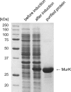
Purity of recombinant MurK analyzed by SDS-PAGE and Coomassie brilliant blue staining. The protein was overproduced in E. coli BL21(DE3) carrying pMurK. Lane 1, protein size standard; lane 2, E. coli cell extract before induction of MurK; lane 3, E. coli cell extract after induction of MurK; lane 4, 20 μg of purified MurK.
Fig. 3.
Fig. 3.
Time course of phosphorylation of GlcNAc (A) and MurNAc (B) by MurK, respectively, yielding GlcNAc-6-phosphate and MurNAc-6-phosphate. ATP was used as phosphate donor in a 2-fold access relative to the amino sugar (250 nmol per assay). The amino sugars were separated by TLC as described in Materials and Methods.
Fig. 4.
Fig. 4.
Identification of the products of the MurK reaction. The products were identified as GlcNAc-6-phosphate and MurNAc-6-phosphate by treatment with the lactyl etherase MurQ, which interconverts MurNAc-6-phosphate and GlcNAc-6-phosphate, yielding an equilibrium of the reaction intermediate Δ-2,3-GlcNAc-6-phosphate and GlcNAc-6-phosphate of 30 to 70%.
Fig. 5.
Fig. 5.
Substrate specificity of MurK. Chemical structures of selected amino sugars and sugars (A) that were tested as substrates for MurK by TLC with nonradioactive (B) and radioactive (C) kinase assays. Spots that can be assigned to phosphorylated sugars are only visual for GlcNAc and MurNAc upon incubation with MurK. Neither anhMurNAc, the disaccharide GlcNAc-MurNAc, nor the muramyl dipeptide (MDP) served as a substrate for MurK. After cleavage of GlcNAc-MurNAc disaccharide with N-acetylglucosaminidase NagZBs the products could be phosphorylated.
Fig. 6.
Fig. 6.
Activity-pH profile of MurK with GlcNAc substrate. A mixture of radioactively labeled and nonradioactive ATP was applied as described in Materials and Methods, and the enzyme activity was quantified by applied TLC. The data are means of three independent experiments; the standard errors are indicated.
Fig. 7.
Fig. 7.
Assay of MurNAc and GlcNAc in cell wall preparation from E. coli. The cell wall sugars can be assayed only after degradation of the peptidoglycan with mutanolysin, AmiD, and/or NagZBs. The monosaccharides GlcNAc and MurNAc were phosphorylated by MurK with [γ-32P]ATP.

Similar articles

Cited by

References

    1. Atrih A., Bacher G., Allmaier G., Williamson M. P., Foster S. J. 1999. Analysis of peptidoglycan structure from vegetative cells of Bacillus subtilis 168 and role of PBP 5 in peptidoglycan maturation. J. Bacteriol. 181:3956–3966 - PMC - PubMed
    1. Bethesda Research Laboratories 1986. BRL pUC host: Escherichia coli DH5α competent cells. Focus 8:9
    1. Blattner F. R., et al. 1997. The complete genome sequence of Escherichia coli K-12. Science 277:1453–1462 - PubMed
    1. Bradford M. M. 1976. A rapid and sensitive method for the quantitation of microgram quantities of protein utilizing the principle of protein-dye binding. Anal. Biochem. 72:248–254 - PubMed
    1. Calandra G. B., Cole R. M. 1980. Lysis and protoplast formation of group B streptococci by mutanolysin. Infect. Immun. 28:1033–1037 - PMC - PubMed

Publication types

MeSH terms

LinkOut - more resources