. 2011 Aug 9;108(32):12996-3001.
doi: 10.1073/pnas.1106379108. Epub 2011 Jul 22.

Combinatorial synthesis of chemically diverse core-shell nanoparticles for intracellular delivery

Affiliations

Combinatorial synthesis of chemically diverse core-shell nanoparticles for intracellular delivery

Daniel J Siegwart et al. Proc Natl Acad Sci U S A. .

Abstract

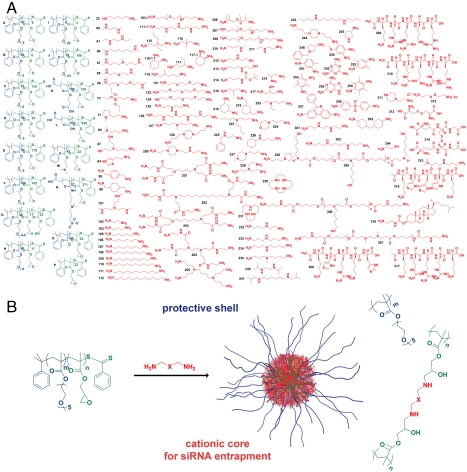
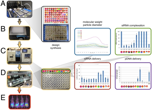
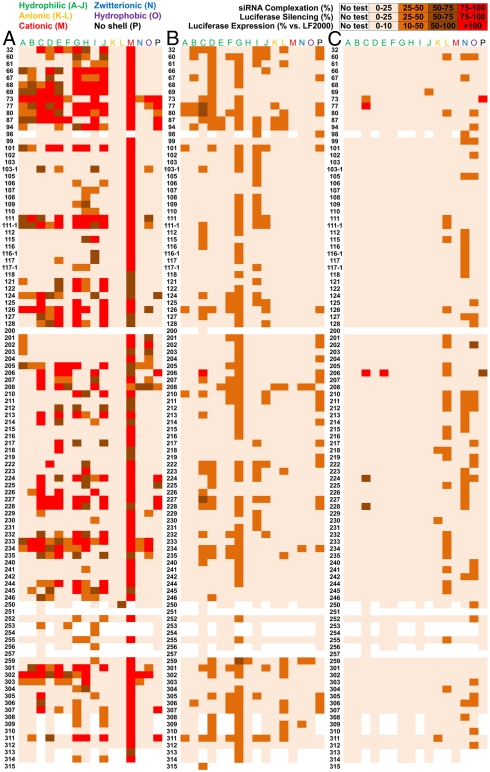
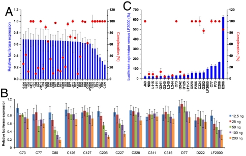
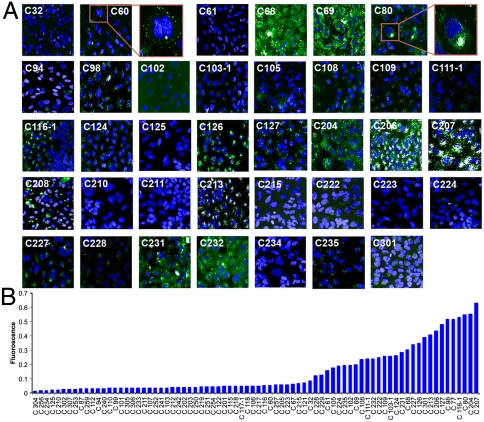
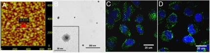
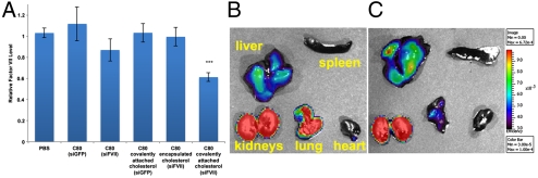
Analogous to an assembly line, we employed a modular design for the high-throughput study of 1,536 structurally distinct nanoparticles with cationic cores and variable shells. This enabled elucidation of complexation, internalization, and delivery trends that could only be learned through evaluation of a large library. Using robotic automation, epoxide-functionalized block polymers were combinatorially cross-linked with a diverse library of amines, followed by measurement of molecular weight, diameter, RNA complexation, cellular internalization, and in vitro siRNA and pDNA delivery. Analysis revealed structure-function relationships and beneficial design guidelines, including a higher reactive block weight fraction, stoichiometric equivalence between epoxides and amines, and thin hydrophilic shells. Cross-linkers optimally possessed tertiary dimethylamine or piperazine groups and potential buffering capacity. Covalent cholesterol attachment allowed for transfection in vivo to liver hepatocytes in mice. The ability to tune the chemical nature of the core and shell may afford utility of these materials in additional applications.


Conflict of interest statement

Conflict of interest statement: R.L. is a shareholder and member of the Scientific Advisory Board of Alnylam. D.G.A. is a consultant with Alnylam. R.L and D.G.A have sponsored research grants from Alnylam. Alnylam also has a license to certain intellectual property invented at MIT. W.Q., C.Z., and T.N. are employed by Alnylam.

Figures

Fig. 1.
Fig. 1.
Combinatorial synthesis of core-shell nanoparticles. (A) Epoxide-containing block copolymers and amines were used to synthesize a combinatorial library of hairy core-shell nanoparticles. (B) Synthesis occurs via the ring-opening reaction of epoxides by amines forming β-hydroxyl groups and cross-linking points separated by the functionality (X) contained in the amine. Each nanoparticle is named by a letter corresponding to the starting block copolymer and a number corresponding to an amine.
Fig. 2.
Fig. 2.
Modular design for the synthesis, characterization, and screening of a library of core-shell nanoparticles. (A) The synthesis of 1,536 core-shell nanoparticles was carried out on a Symyx fluid-handling robot inside of glass vials in a 96-well plate format. (B) The nanoparticles were purified and filtered through a HT filtering apparatus into a standard 96-well tissue culture plate. (C) The MW and particle size of the polymers were measured by a HT GPC system. (D) The same plate format was used for an RNA complexation assay, carried out on a Tecan cell culture robot. HT in vitro screens for siRNA and pDNA delivery were performed. (E) Biodistribution of C227 (left two images) and C80 (right two images) are shown. GPC, siRNA complexation data, and in vitro siRNA and pDNA delivery results with standard deviation (s.d.) for nanoparticles C32-C94 (positions A1-A12 in the plate) are presented.
Fig. 3.
Fig. 3.
In vitro screening of core-shell nanoparticles. (A) The degree of RNA entrapment was quantified using the RiboGreen assay. The average percent of complexation is shown. (B) HeLa cells stably expressing both firefly and Renilla luciferases were treated with firefly targeting siRNA-nanoparticles complexes. The average percent reduction in firefly luciferase activity after treatment is shown. (C) HeLa cells were treated with pDNA(luc)-nanoparticle complexes. Activity using LF2000 was standardized to 100. The average percent activity versus LF2000 after treatment is shown. To provide a picture of the capability of all nanoparticles, the complexation, siRNA delivery, and pDNA experiments were performed in triplicate at a weight ratio of 50∶1 (nanoparticle:siRNA).
Fig. 4.
Fig. 4.
In vitro core-shell nanoparticle-mediated delivery trends. (A) RNA complexation and luciferase silencing after delivery of nanoparticle:siRNA complexes (50∶1, wt/wt) is expressed for the top performing nanoparticles. (B) siRNA dose response for the top nanoparticles. (C) Complexation and luciferase activity after delivery of nanoparticle:pDNA complexes (50∶1, wt/wt) is expressed for the top performing nanoparticles. (AC) n = 4; s.d. is expressed.
Fig. 5.
Fig. 5.
In vitro uptake screening. (A) Cellular internalization of selected nanoparticles after 1 hr of incubation is demonstrated by HT automated confocal microscopy. HeLa cells were exposed to all C-based nanoparticles complexed with Alexa-594 siRNA (50∶1, wt/wt). Twenty different fields were imaged and a representative image of nanoparticle/siRNA complex is presented (Alexa 594 is pseudocolored green). Additional images appear in Fig. S4. (B) The total fluorescence was quantified and plotted. False positives were identified by visible aggregation (e.g., C208 included for reference) or significant background and were removed from the graph. Interestingly, C60 nanoparticles interact with the cell membrane but did not enter cells, while C80 nanoparticles were effectively internalized (see zoom insets).
Fig. 6.
Fig. 6.
Characterization of core-shell nanoparticles. (A) Dried FF110 nanoparticles on mica showed uniform particle size around 34 nm by AFM. (B) TEM was utilized to visualize the core-shell structure of R126 nanoparticles. The insert clearly showed an electron dense cross-linked core, and a more electron loose shell. These TEM images are unstained. Additional AFM and TEM images appear in Fig. S3. Cell internalization of FITC-C80 (C) and FITC-C227 (D) nanoparticles after 1 hr of incubation was demonstrated by confocal microscopy. Punctate green fluorescence was observed within the cell.
Fig. 7.
Fig. 7.
In vivo silencing of Factor VII in liver hepatocytes. Nanoparticles were purified by dialysis, complexed with siRNA at a weight ratio of 10∶1 (nanoparticle:siRNA), and delivered intravenously to C57BL/6 mice. Mice received a single bolus administration of 5 mg/kg total siRNA via tail-vein injection and Factor VII levels were quantified 48 hr post injection. (A) Covalent attachment of cholesterol to C80 nanoparticles enabled silencing. Noncovalent encapsulation of cholesterol and delivery of control siGFP resulted in no knockdown. Data points represent group mean ± s.d. Data points marked with asterisks are significant relative to control treated groups (***, P < 0.0001; t-test, double-tailed, n = 14). Representative images of isolated organs for Cy5-siRNA-C80 (b) and Cy5-siRNA-C80-10 mol% cholesterol (C) nanoparticle complexes demonstrated nanoparticle accumulation in the liver, kidneys, and lungs after 120 min. The covalent attachment of cholesterol to C80 resulted in enhanced liver accumulation and reduced lung accumulation.

References

    1. Balazs A, Emrick T, Russell T. Nanoparticle polymer composites: Where two small worlds meet. Science. 2006;314:1107–1110. - PubMed
    1. Taton T, Mirkin C, Letsinger R. Scanometric DNA array detection with nanoparticle probes. Science. 2000;289:1757–1760. - PubMed
    1. Shipway A, Katz E, Willner I. Nanoparticle arrays on surfaces for electronic, optical, and sensor applications. ChemPhysChem. 2000;1:18–52. - PubMed
    1. LaVan D, Lynn D, Langer R. Moving smaller in drug discovery and delivery. Nat Rev Drug Discov. 2002;1:77–84. - PubMed
    1. Davis ME, Chen Z, Shin DM. Nanoparticle therapeutics: An emerging treatment modality for cancer. Nat Rev Drug Discov. 2008;7(9):771–782. - PubMed

Publication types

LinkOut - more resources