. 2011 Aug 9;108(32):13077-82.
doi: 10.1073/pnas.1107719108. Epub 2011 Jul 22.

Tapping natural reservoirs of homing endonucleases for targeted gene modification

Affiliations

Tapping natural reservoirs of homing endonucleases for targeted gene modification

Ryo Takeuchi et al. Proc Natl Acad Sci U S A. .

Abstract

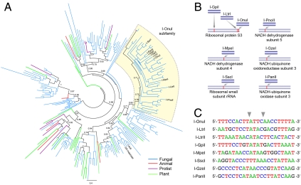
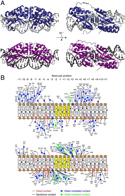
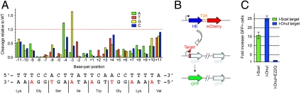
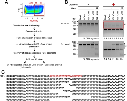
Homing endonucleases mobilize their own genes by generating double-strand breaks at individual target sites within potential host DNA. Because of their high specificity, these proteins are used for "genome editing" in higher eukaryotes. However, alteration of homing endonuclease specificity is quite challenging. Here we describe the identification and phylogenetic analysis of over 200 naturally occurring LAGLIDADG homing endonucleases (LHEs). Biochemical and structural characterization of endonucleases from one clade within the phylogenetic tree demonstrates strong conservation of protein structure contrasted against highly diverged DNA target sites and indicates that a significant fraction of these proteins are sufficiently stable and active to serve as engineering scaffolds. This information was exploited to create a targeting enzyme to disrupt the endogenous monoamine oxidase B gene in human cells. The ubiquitous presence and diversity of LHEs described in this study may facilitate the creation of many tailored nucleases for genome editing.


Conflict of interest statement

Conflict of interest statement: B.L.S. and A.M.S. are founders of a biotechnology company (Precision Genome Engineering) that conducts engineering and selection studies on homing endonucleases for gene targeting.

Figures

Fig. 1.
Fig. 1.
Diversity of LHE genes and their sequence recognition specificities. (A) Shown is a phylogenetic analysis of 211 single-chain LHEs as identified by structure-based alignments. Branches are color-coded according to the taxonomic source from which the LHE was identified, as indicated. Groups of LHEs are colored the same if all members of that subfamily are derived from the same taxonomic group. Black lines indicate that members of a subfamily are derived from more than one taxonomic grouping. The I-OnuI subfamily is highlighted in yellow, with individual LHEs named (e.g., I-OnuI). The phylogenetic tree was generated by PhyML (14), and approximate likelihood-ratio test values greater than 0.7 are indicated on major nodes. A version of the tree with branches labeled by accession numbers and partial sequences of all the LHEs identified are provided as Fig. S1A and Dataset S1, respectively. (B) Schematic of homing sites recognized by the I-OnuI subfamily. (C) Target sequences for I-OnuI, I-LtrI, and I-LtrII were identified in previous studies (13, 15). Recognition sequences for the other LHEs were predicted through comparative sequence alignments of each host gene to related species lacking an embedded endonuclease. Cleavage activity against each predicted site was verified using yeast surface-displayed enzyme in both in vitro and flow cytometric cleavage assays (see SI Text for detail). Sequence analysis of cleaved products showed that all of the homologues generated 3′, 4-base overhangs by hydrolyzing the phosphodiester bonds between the base-pair positions ± 2 and ± 3 (indicated by gray arrows).
Fig. 2.
Fig. 2.
Structure determination of I-OnuI and I-LtrI. (A) Crystal structures of I-OnuI (Upper, dark blue) and I-LtrI (Lower, purple) bound to their physiological target sites. Residues 156–158 in the middle of linker between the two pseudosymmetric half domains of I-OnuI were disordered (represented as black dots). The loop region (residues 236–244) between the third and fourth β-sheets of the C-terminal half domain of I-LtrI could not be assigned due to poor electron density. (B) Schematic of I-OnuI (Upper) and I-LtrI (Lower) DNA contacts. The two scissile phosphates and the other backbone phosphates are depicted as dark blue and orange spheres, respectively. The central four base pairs (positions ± 1 and ± 2) are colored in yellow. Residue numbers of I-LtrI crystal shown here are shifted from the numbers assigned in the deposited PDB file, in order to align the residue numbers of the first LAGLIDADG motif to the corresponding numbers of I-OnuI crystal. E29 in the original PDB file is labeled as E22.
Fig. 3.
Fig. 3.
Evaluation of specificity and activity of I-OnuI. (A) Cleavage activity of I-OnuI for each single base-pair substituted target site from its physiological target was assayed by tethering fluorescence-labeled substrates to the I-OnuI protein expressed on the surface of yeast. Each bar represents relative cleavage activity for a target site containing a single base-pair substitution from the WT I-OnuI target. A, green; T, red; G, yellow; C, blue. The bottom strand encodes a host gene product, and wobble positions in the host gene reading frame are colored in red. (B) Schematic representation of the plasmids used in an episomal gene conversion assay. A homing endonuclease (HE) gene was linked to the mCherry gene through a 2A peptide sequence from T2A, leading to a coexpression of the two genes separated by the ribosomal skipping mechanism (23). The episomal DR-GFP reporter harbored two nonfunctional GFP gene (24): One was interrupted by a LHE target site and a stop codon, and the other was truncated. Double-strand breaks induced by a HE promote the conversion between the two nonfunctional GFP genes on the episomal DR-GFP reporter, resulting in the restoration of the GFP expression. (C) Gene conversion activity was assayed using the episomal DR-GFP reporter containing a target site for a LHE. Each bar represents an increase in a fraction of GFP positive cells by transient expression of a LHE compared to the background observed by transfection with the DR-GFP reporter alone. Errors refer to ± SD of three independent experiments.
Fig. 4.
Fig. 4.
Redesign of I-OnuI to target the human monoamine oxidase B gene. (A) Sequence alignment of the I-OnuI and MAO-B target sites (Upper) or the human MAO-B target and the corresponding sequences coded in other mammalian genomes (Lower). (B) An episomal GFP gene conversion assay was carried out similar to those shown in Fig. 3C. Error bars refer to ± SD of three independent experiments. E1 I-OnuI was selected from directed evolution of I-OnuI in bacteria, and E2 I-OnuI was generated by an addition of E178D in E1 I-OnuI. (C) In vitro cleavage activity was assayed using 32P-labeled DNA substrates. Error bars refer to ± SD of three independent experiments. The I-OnuI target is shown as closed squares and the MAO-B target as open squares; I-OnuI, black; E1 I-OnuI, blue; E2 I-OnuI, red. (D) Cleavage activity of E2 I-OnuI for a target sequence containing a single base-pair substitution from the MAO-B target was tested similarly to that shown in Fig. 3A. A, green; T, red; G, yellow; C, blue.
Fig. 5.
Fig. 5.
Targeted mutagenesis of the endogenous MAO-B gene in human tissue culture cells. (A) Experimental procedure for detection of mutations at the endogenous MAO-B gene locus that are induced by transiently expressed I-OnuI variants. The sorting gates H and M were set to collect approximately the top 25% and the following 25% of mCherry positive cells. The mCherry is cotranslated with I-OnuI variants as a separate protein (Fig. 3B). (B) The impaired, chromosomal MAO-B target was detected by in vitro digestion with E2 I-OnuI recombinant protein, following PCR amplification of the surrounding sequence. Asterisks indicate cleavage-resistant (CR) fragments that are significantly or selectively observed in PCR amplicons from E1 or E2 I-OnuI-transfected cells. DNAs larger than the cleaved fragments in the first round of digestion (included in a red box; Upper Right) were recovered, reamplified, and subjected to the second round of digestion (Lower Panel). (C) CR fragments from E2 I-OnuI-transfected cells (collected in the sorting gate H) were analyzed by sequencing. Small deletions were found within the endogenous MAO-B target site. The intact genome sequence is shown on the top (WT), and the MAO-B target site is in red.

References

    1. Christian M, et al. Targeting DNA double-strand breaks with TAL effector nucleases. Genetics. 2010;186:757–761. - PMC - PubMed
    1. Li T, et al. TAL nucleases (TALNs): Hybrid proteins composed of TAL effectors and FokI DNA-cleavage domain. Nucleic Acids Res. 2010;39:359–372. - PMC - PubMed
    1. Marcaida MJ, Munoz IG, Blanco FJ, Prieto J, Montoya G. Homing endonucleases: from basics to therapeutic applications. Cell Mol Life Sci. 2010;67:727–748. - PMC - PubMed
    1. Stoddard BL. Homing endonucleases: From microbial genetic invaders to reagents for targeted DNA modification. Structure. 2011;19:7–15. - PMC - PubMed
    1. Urnov FD, Rebar EJ, Holmes MC, Zhang HS, Gregory PD. Genome editing with engineered zinc finger nucleases. Nat Rev Genet. 2010;11:636–646. - PubMed

Publication types

LinkOut - more resources