Review
. 2011 Oct;301(4):H1220-8.
doi: 10.1152/ajpheart.00601.2011. Epub 2011 Jul 22.

Desmin-related cardiomyopathy: an unfolding story

Affiliations
Review

Desmin-related cardiomyopathy: an unfolding story

Patrick M McLendon et al. Am J Physiol Heart Circ Physiol. 2011 Oct.

Abstract

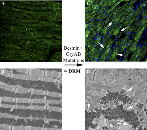
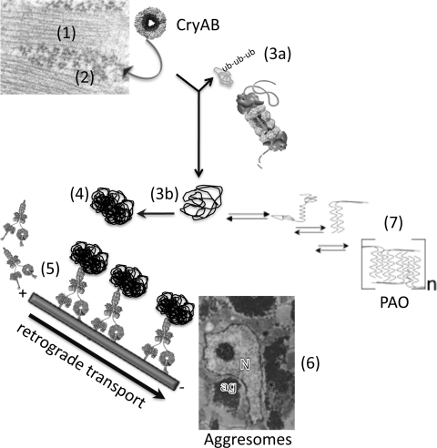
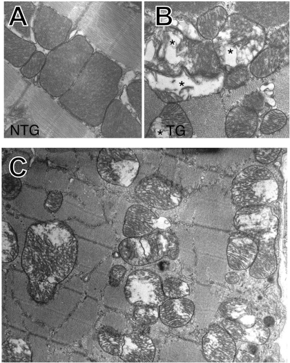
The intermediate filament protein desmin is an integral component of the cardiomyocyte and serves to maintain the overall structure and cytoskeletal organization within striated muscle cells. Desmin-related myopathy can be caused by mutations in desmin or associated proteins, which leads to intracellular accumulation of misfolded protein and production of soluble pre-amyloid oligomers, which leads to weakened skeletal and cardiac muscle. In this review, we examine the cellular phenotypes in relevant animal models of desmin-related cardiomyopathy. These models display characteristic sarcoplasmic protein aggregates. Aberrant protein aggregation leads to mitochondrial dysfunction, abnormal metabolism, and altered cardiomyocyte structure. These deficits to cardiomyocyte function may stem from impaired cellular proteolytic mechanisms. The data obtained from these models allow a more complete picture of the pathology in desmin-related cardiomyopathy to be described. Moreover, these studies highlight the importance of desmin in maintaining cardiomyocyte structure and illustrate how disrupting this network can be deleterious to the heart. We emphasize the similarities observed between desmin-related cardiomyopathy and other protein conformational disorders and speculate that therapies to treat this disease may be broadly applicable to diverse protein aggregation-based disorders.


Figures

Fig. 1.
Fig. 1.
Desmin and αB-crystallin (CryAB) mutations cause desmin-related (cardio)myopathy (DRM). Immunofluorescent staining with anti-desmin (A and C) is detected by the green color. A and B: heart tissue from a nontransgenic (NTG) mouse. A: immunohistochemical staining for desmin. The intercalated disks are apparent. B: normal heart tissue contains regular sarcomeres (B) that exhibit a striated morphology with well-ordered mitochondria regularly positioned about the sarcomeres. C and D: mutations in desmin and CryAB disrupt the desmin cytoskeletal network, which perturbs normal organization of the myofibrils. These examples are of heart tissue from a DesD7 transgenic (TG) mouse. C: fluorescence micrograph showing sarcomere disruption and intracellular protein aggregates (white arrows) that positively stain for desmin. D: electron micrograph showing sarcomeric disarray, electron-dense aggregates (white arrows), and interrupted mitochondrial positioning about the sarcomere. The sarcomeric disorganization and aggregate formation are common phenotypes of DRM at the cellular level.
Fig. 2.
Fig. 2.
Genesis of aggresomes and pre-amyloid oligomers (PAO) in cardiomyocytes expressing mutant CryAB. Shown in schematic form are the processes that can lead to generation of the perinuclear aggregates or aggresomes and PAO in cardiomyocytes expressing CryABR120G. 1: Shown attached to a nascent sarcomere are polysomes (2) in the process of making sarcomeric proteins. Oligomers of mutant CryAB attach to the nascent polypeptide but are unable to mediate normal folding. 3a: Misfolded protein is recognized as such, ubiquitinated (ub-ub-ub) and targeted for degradation by the proteasome. The protein is degraded and the components recycled. 3b and 4: The misfolded protein, either because its size precludes it from entering the bore of the proteasome or because of compromised proteasome function, is not degraded and continues to form larger aggregates. 5: The aggregates are attached to dynein motors and are retrogradely transported down the microtubules to a perinuclear location. 6: These coalesce and form aggresomes, which are shown as electron dense, granulofilamentous aggregates near the nucleus (Ref. 58). N, nucleus; ag, aggresome. 7: It has been estimated that ∼3,000 proteins have amyloidogenic potential. Under favorable circumstances, for those proteins, they can assume a parallel, β-sheet structure and form soluble PAO of indeterminate number (n). While the precise cause-and-effect relationships between the observed cellular phenotypes and corresponding cellular dysfunction are not clear, mitochondrial disruption, increased oxidative stress, and cardiomyocyte death can all result from the observed deficits in chaperone function, proteolytic activity, and PAO/aggregate generation.
Fig. 3.
Fig. 3.
Expression of CryABR120G results in mitochondrial pathology. A: a high-magnification, electron-microscopy picture of a normal mouse cardiac sarcomere at 8 wk postbirth, showing the defined organization of a dyad of mitochondria juxtaposed over the A and I bands of the sarcomere. B: a picture from an 8-wk-old CryABR120G heart showing the characteristic altered mitochondria organization and swollen, lysed mitochondria as well. C: a larger field showing the frequency of the cristolysis and mitochondrial swelling and disorganization relative to one another and the underlying sarcomeres. *Areas showing significant mitochondrial cristolysis.

References

    1. Bär H, Strelkov SV, Sjöberg G, Aebi U, Herrmann H. The biology of desmin filaments: how do mutations affect their structure, assembly, and organisation? J Struct Biol 148: 137–152, 2004 - PubMed
    1. Bence NF, Sampat RM, Kopito RR. Impairment of the ubiquitin-proteasome system by protein aggregation. Science 292: 1552–1555, 2001 - PubMed
    1. Bova MP, Yaron O, Huang Q, Ding L, Haley DA, Stewart PL, Horwitz J. Mutation R120G in αB-crystallin, which is linked to a desmin-related myopathy, results in an irregular structure and defective chaperone-like function. Proc Natl Acad Sci USA 96: 6137–6142, 1999 - PMC - PubMed
    1. Brady JP, Garland DL, Green DE, Tamm ER, Giblin FJ, Wawrousek EF. αB-crystallin in lens development and muscle integrity: a gene knockout approach. Invest Ophthalmol Vis Sci 42: 2924–2934, 2001 - PubMed
    1. Capetanaki Y. Desmin cytoskeleton: a potential regulator of muscle mitochondrial behavior and function. Trends Cardiovasc Med 12: 339–348, 2002 - PubMed

Publication types