. 2011 Aug;121(8):3042-50.
doi: 10.1172/JCI46276. Epub 2011 Jul 25.

Prolactin increases SMN expression and survival in a mouse model of severe spinal muscular atrophy via the STAT5 pathway

Affiliations

Prolactin increases SMN expression and survival in a mouse model of severe spinal muscular atrophy via the STAT5 pathway

Faraz Farooq et al. J Clin Invest. 2011 Aug.

Erratum in

  • J Clin Invest. 2011 Sep 1;121(9):3763

Abstract

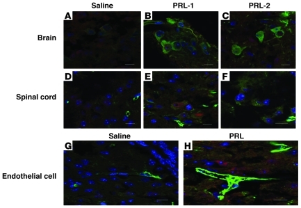
Spinal muscular atrophy (SMA) is an autosomal recessive neurodegenerative disease that is characterized by the loss of motor neurons, resulting in progressive muscle atrophy. It is caused by the loss of functional survival motor neuron (SMN) protein due to mutations or deletion in the SMN1 gene. A potential treatment strategy for SMA is to upregulate levels of SMN protein. Several agents that activate STAT5 in human and mouse cell lines enhance SMN expression from the SMN2 gene and can compensate, at least in part, for the loss of production of a functional protein from SMN1. Here, we have shown that prolactin (PRL) increases SMN levels via activation of the STAT5 pathway. PRL increased SMN mRNA and protein levels in cultured human and mouse neuronal cells. Administration of STAT5-specific siRNA blocked the effects of PRL, indicating that the PRL-induced transcriptional upregulation of the SMN-encoding gene was mediated by activation of STAT5. Furthermore, systemic administration of PRL to WT mice induced SMN expression in the brain and spinal cord. Critically, PRL treatment increased SMN levels, improved motor function, and enhanced survival in a mouse model of severe SMA. Our results confirm earlier work suggesting STAT5 pathway activators as potential therapeutic compounds for the treatment of SMA and identify PRL as one such promising agent.


Figures

Figure 1
Figure 1. PRL treatment upregulates SMN mRNA and protein in vitro.
NT2 or MN-1 cells were treated with PRL (0, 25, 50, and 100 ng/ml) and then harvested at indicated intervals for RT-PCR or Western blot analyses. (A) Presence of PRLR in NT2 cells. (B) Quantification of SMN mRNA relative to β-actin in NT2 cells after PRL treatment (fold induction; the ratio at 0 hour was set as 1). Mean + SD of 3 independent experiments. (C) Representative Western blot showing effect of PRL on SMN protein in NT2 cells. (D) Densitometric quantification of SMN relative to β-actin is shown for NT2 cells. Mean + SD of 3 independent experiments. (E) Representative Western blot showing effect of PRL on SMN protein in MN-1 cells. (F) Densitometric quantifications of Smn relative to tubulin are shown for MN-1 cells. Mean + SD of 3 independent experiments.
Figure 2
Figure 2. PRL increases SMN expression via STAT5 pathway.
(A) Representative Western blot showing activation of STAT5 pathway upon PRL treatment in NT2 cells. (B) ATA (STAT5 pathway activator) treatment upregulates SMN mRNA in vitro. NT2 cells were treated with ATA (250 μM) and then harvested after 48 hours for RT-PCR. Quantification of SMN mRNA relative to β-actin in NT2 cells after ATA treatment (fold induction; the ratio at 0 hours was set as 1). Mean + SD of 3 independent experiments is shown. (C) The siRNA knockdown of STAT5 protein attenuates PRL-induced increase in SMN protein. Representative Western blots showing both STAT5 knockdown and its effect on PRL-induced increase in SMN protein. s. seq., scrambled sequence. (D) Densitometric quantification of SMN relative to tubulin is shown. Mean + SD of 3 independent experiments is shown.
Figure 3
Figure 3. PRL upregulates Smn protein in WT mice.
Male CD-1 WT mice were treated daily with PRL (5.0, 50.0, 500.0 μg/kg) for 5 days, then sacrificed. Brain and spinal cord tissues were harvested for Western blot analysis. (A) Representative Western blot showing the effect of PRL on Smn protein in brain samples of CD-1 mice treated with saline (control, lane 1) or PRL (lanes 2, 3, and 4, respectively) (n = 3). (B) Densitometric quantification of Smn relative to tubulin is shown for brain samples. (C) Representative Western blot showing the effect of PRL on Smn protein in spinal cord samples of CD-1 mice treated with saline (control, lane 1) or PRL (lanes 2, 3, and 4) (n = 3). (D) Densitometric quantification of Smn relative to tubulin is shown for spinal cord samples. Mean + SD.
Figure 4
Figure 4. PRL upregulates Smn protein in SMA mouse model.
SMAΔ7 mice were treated daily with saline or PRL (2.5 mg/kg) from P1 for 6 days, then sacrificed at P7. Brain and spinal cord tissues were harvested for RT-PCR and Western blot analysis. (A) Quantification of Smn mRNA relative to β-actin in brain tissue after PRL treatment (fold induction; the ratio at saline treatment was set as 1). Mean + SD is shown (n = 5). (B) Quantification of Smn mRNA relative to β-actin in spinal cord tissue after PRL treatment (fold induction; the ratio at saline treatment was set as 1). Mean + SD is shown (n = 5). (C) Representative Western blot showing effect of PRL on Smn protein in brain samples of SMAΔ7 mice treated with saline (control, lanes 1, 2, and 3) or PRL (lanes 4, 5, and 6, respectively) (each lane represents individual animal; all lanes were run on the same gel but were noncontiguous). (D) Densitometric quantification of SMN relative to tubulin is shown for brain samples. Mean + SD. (E) Representative Western blot showing effect of PRL on Smn protein in spinal cord samples of SMAΔ7 mice treated with saline (control, lanes 1, 2, and 3) or PRL (lanes 4, 5, and 6, respectively) (each lane represents individual animal; all lanes were run on the same gel but were noncontiguous). (F) Densitometric quantification of SMN relative to tubulin is shown for spinal cord samples. Mean + SD.
Figure 5
Figure 5. PRL upregulates Smn protein expression in motor neurons and endothelial cells in SMA mouse model.
SMAΔ7 mice were treated daily with saline or PRL (2.5 mg/kg) from P1 for 6 days, then sacrificed at P7. Brain stem and spinal cord tissues were harvested for immunohistochemistry analysis. Representative merged confocal images (Smn/Alexa Fluor 488 [green] + HB9/Alexa Fluor 568 [red; motor neuron marker] plus Hoechst [blue]) for different tissues are shown. Representative confocal images showing effect of PRL on Smn protein expression in brain stem and spinal cord motor neurons samples of SMAΔ7 mice treated with saline (control, A and D) or PRL (B and C, and E and F), respectively (n = 3). Scale bars: 10 μM. Representative confocal images showing effect of PRL on Smn protein expression in endothelial cells of SMAΔ7 mice treated with saline (control, G) or PRL (H) (n = 3). Scale bars: 10 μM.
Figure 6
Figure 6. PRL ameliorates disease phenotype in SMA mouse model.
SMAΔ7 mice were treated daily with i.p. injections of PRL (2.5 mg/kg) from P1 onward. (A) Weights of SMAΔ7 mice treated with PRL (squares) or saline (circles) (n = 5) and weights for heterozygous mice treated with saline (triangles) are also shown for comparison. Mean ± SD. (B) Righting times of SMAΔ7 mice treated with PRL (squares) or saline (circles) (n = 5). Mean ± SD. (C) Kaplan-Meier survival curves of SMAΔ7 mice treated with PRL (squares) or vehicle (circles) (n = 10); ***P < 0.0001, log-rank test.
Figure 7
Figure 7. Smn protein level is upregulated in SMAΔ7 mice until time of death.
SMAΔ7 mice were treated daily with PRL (2.5 mg/kg) from P1 onward. Brain, spinal cord, and muscle tissues were harvested upon death for Western blot analysis. (A) Representative Western blot showing the effect of PRL on Smn protein in brain samples of SMAΔ7 mice treated with saline (control, lane 1, 2, and 3) or PRL (lane 4, 5, 6, and 7) (each lane represents individual animal; all lanes were run on the same gel but were noncontiguous). (B) Densitometric quantification of SMN relative to tubulin is shown for brain samples. Mean ± SD. (C) Representative Western blot showing effect of PRL on Smn protein in spinal cord samples of SMAΔ7 mice treated with saline (control, lanes 1, 2, and 3) or PRL (lanes 4, 5, 6, and 7, respectively) (each lane represents individual animal). (D) Densitometric quantification of SMN relative to tubulin is shown for spinal cord samples. Mean ± SD. (E) Representative Western blot showing the effect of PRL on Smn protein in muscle samples of SMAΔ7 mice treated with saline (control, lanes 1, 2, and 3) or PRL (lanes 4, 5, 6, and 7) (each lane represents individual animal). (F) Densitometric quantification of SMN relative to tubulin is shown for muscle samples. Mean ± SD.
Figure 8
Figure 8. Proposed model for PRL-mediated induction of SMN in motor neurons.
PRL treatment causes phosphorylation and activation of STAT5, pathway which results in transcriptional upregulation of SMN gene, resulting in an increase in SMN mRNA levels. This ultimately increases SMN protein expression.

Comment in

References

    1. Pearn J. Incidence, prevalence, and gene frequency studies of chronic childhood spinal muscular atrophy. J Med Genet. 1978;15(6):409–413. doi: 10.1136/jmg.15.6.409. - DOI - PMC - PubMed
    1. Emery AE. Population frequencies of inherited neuromuscular diseases--a world survey. Neuromuscul Disord. 1991;1(1):19–29. doi: 10.1016/0960-8966(91)90039-U. - DOI - PubMed
    1. Lefebvre S, et al. Identification and characterization of a spinal muscular atrophy-determining gene. Cell. 1995;80(1):155–165. doi: 10.1016/0092-8674(95)90460-3. - DOI - PubMed
    1. Friesen WJ, Dreyfuss G. Specific sequences of the Sm and Sm-like (Lsm) proteins mediate their interaction with the spinal muscular atrophy disease gene product (SMN). J Biol Chem. 2000;275(34):26370–26375. doi: 10.1074/jbc.M003299200. - DOI - PubMed
    1. Buhler D, Raker V, Luhrmann R, Fischer U. Essential role for the tudor domain of SMN in spliceosomal U snRNP assembly: implications for spinal muscular atrophy. Hum Mol Genet. 1999;8(13):2351–2357. doi: 10.1093/hmg/8.13.2351. - DOI - PubMed

Publication types