

Tumor-selective, adenoviral-mediated GFP genetic labeling of human cancer in the live mouse reports future recurrence after resection
- PMID: 21785265
- PMCID: PMC3219541
- DOI: 10.4161/cc.10.16.16756
Tumor-selective, adenoviral-mediated GFP genetic labeling of human cancer in the live mouse reports future recurrence after resection
Abstract
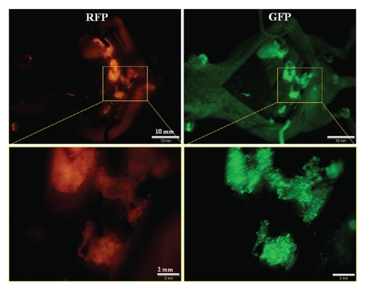
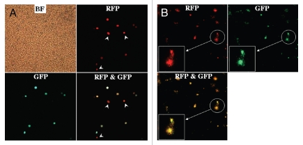
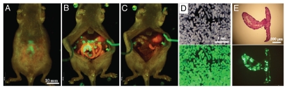
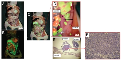
We have previously developed a telomerase-specific replicating adenovirus expressing GFP (OBP-401), which can selectively label tumors in vivo with GFP. Intraperitoneal (i.p.) injection of OBP-401 specifically labeled peritoneal tumors with GFP, enabling fluorescence visualization of the disseminated disease and real-time fluorescence surgical navigation. However, the technical problems with removing all cancer cells still remain, even with fluorescence-guided surgery. In this study, we report imaging of tumor recurrence after fluorescence-guided surgery of tumors labeled in vivo with the telomerase-dependent, GFP-containing adenovirus OBP-401.. Recurrent tumor nodules brightly expressed GFP, indicating that initial OBP-401-GFP labeling of peritoneal disease was genetically stable, such that proliferating residual cancer cells still express GFP. In situ tumor labeling with a genetic reporter has important advantages over antibody and other non-genetic labeling of tumors, since residual disease remains labeled during recurrence and can be further resected under fluorescence guidance.
Figures
Similar articles
-
In vivo internal tumor illumination by telomerase-dependent adenoviral GFP for precise surgical navigation.Proc Natl Acad Sci U S A. 2009 Aug 25;106(34):14514-7. doi: 10.1073/pnas.0906388106. Epub 2009 Aug 17. Proc Natl Acad Sci U S A. 2009. PMID: 19706537 Free PMC article.
-
Targeting tumors with a killer-reporter adenovirus for curative fluorescence-guided surgery of soft-tissue sarcoma.Oncotarget. 2015 May 30;6(15):13133-48. doi: 10.18632/oncotarget.3811. Oncotarget. 2015. PMID: 26033451 Free PMC article.
-
Visualization of intrathoracically disseminated solid tumors in mice with optical imaging by telomerase-specific amplification of a transferred green fluorescent protein gene.Cancer Res. 2004 Sep 1;64(17):6259-65. doi: 10.1158/0008-5472.CAN-04-1335. Cancer Res. 2004. PMID: 15342413
-
Diagnostic and therapeutic application of telomerase-specific oncolytic adenoviral agents.Front Biosci. 2008 Jan 1;13:1881-6. doi: 10.2741/2807. Front Biosci. 2008. PMID: 17981675 Review.
-
Imaging the microenvironment of pancreatic cancer patient-derived orthotopic xenografts (PDOX) growing in transgenic nude mice expressing GFP, RFP, or CFP.Cancer Lett. 2016 Sep 28;380(1):349-55. doi: 10.1016/j.canlet.2015.12.021. Epub 2015 Dec 29. Cancer Lett. 2016. PMID: 26742463 Review.
Cited by
-
A topically-sprayable, activatable fluorescent and retaining probe, SPiDER-βGal for detecting cancer: Advantages of anchoring to cellular proteins after activation.Oncotarget. 2017 Jun 13;8(24):39512-39521. doi: 10.18632/oncotarget.17080. Oncotarget. 2017. PMID: 28467810 Free PMC article.
-
Fluorescence endoscopic detection of murine colitis-associated colon cancer by topically applied enzymatically rapid-activatable probe.Gut. 2013 Aug;62(8):1179-86. doi: 10.1136/gutjnl-2011-301795. Epub 2012 Jun 14. Gut. 2013. PMID: 22698650 Free PMC article.
-
Adenoviral targeting of malignant melanoma for fluorescence-guided surgery prevents recurrence in orthotopic nude-mouse models.Oncotarget. 2016 Apr 5;7(14):18558-72. doi: 10.18632/oncotarget.6670. Oncotarget. 2016. PMID: 26701857 Free PMC article.
-
Characteristics of ovarian cancer detection by a near-infrared fluorescent probe activated by human NAD(P)H: quinone oxidoreductase isozyme 1 (hNQO1).Oncotarget. 2017 May 20;8(37):61181-61192. doi: 10.18632/oncotarget.18044. eCollection 2017 Sep 22. Oncotarget. 2017. PMID: 28977855 Free PMC article.
-
Fluorescent Anti-MUC5AC Brightly Targets Pancreatic Cancer in a Patient-derived Orthotopic Xenograft.In Vivo. 2022 Jan-Feb;36(1):57-62. doi: 10.21873/invivo.12676. In Vivo. 2022. PMID: 34972700 Free PMC article.
References
-
- Chishima T, Miyagi Y, Wang X, Yamaoka H, Shimada H, Moossa AR, et al. Cancer invasion and micrometastasis visualized in live tissue by green fluorescent protein expression. Cancer Res. 1997;57:2042–2047. - PubMed
Publication types
MeSH terms
Substances
Grants and funding
LinkOut - more resources
Full Text Sources