. 2011 Jul 22;30(16):3337-52.
doi: 10.1038/emboj.2011.242.

Targeting of Nbp1 to the inner nuclear membrane is essential for spindle pole body duplication

Affiliations

Targeting of Nbp1 to the inner nuclear membrane is essential for spindle pole body duplication

Thomas Kupke et al. EMBO J. .

Abstract

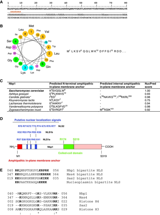
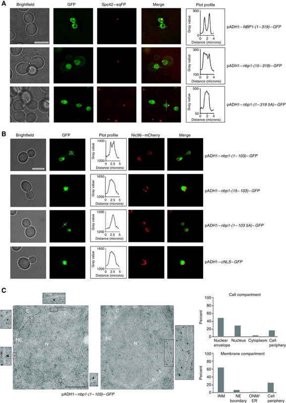
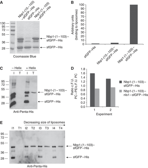
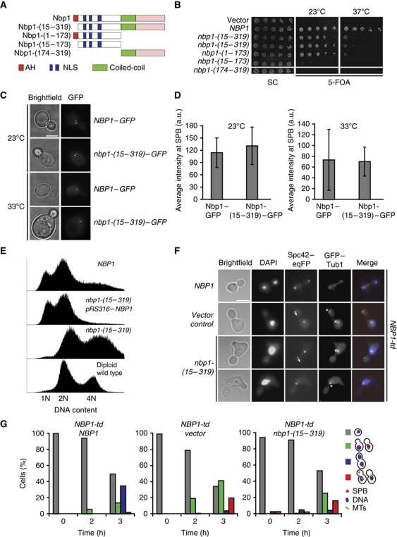
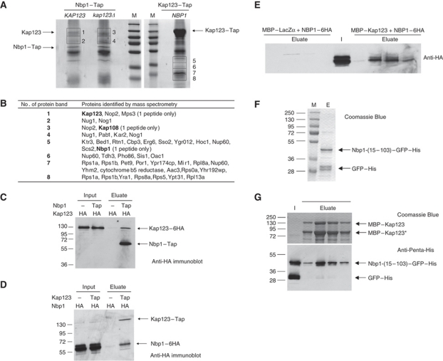
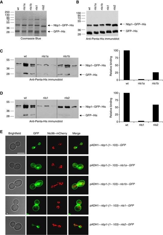
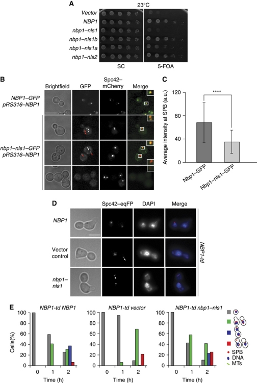
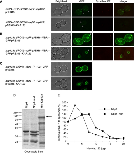
Spindle pole bodies (SPBs), like nuclear pore complexes, are embedded in the nuclear envelope (NE) at sites of fusion of the inner and outer nuclear membranes. A network of interacting proteins is required to insert a cytoplasmic SPB precursor into the NE. A central player of this network is Nbp1 that interacts with the conserved integral membrane protein Ndc1. Here, we establish that Nbp1 is a monotopic membrane protein that is essential for SPB insertion at the inner face of the NE. In vitro and in vivo studies identified an N-terminal amphipathic α-helix of Nbp1 as a membrane-binding element, with crucial functions in SPB duplication. The karyopherin Kap123 binds to a nuclear localization sequence next to this amphipathic α-helix and prevents unspecific tethering of Nbp1 to membranes. After transport into the nucleus, Nbp1 binds to the inner nuclear membrane. These data define the targeting pathway of a SPB component and suggest that the amphipathic α-helix of Nbp1 is important for SPB insertion into the NE from within the nucleus.


Conflict of interest statement

The authors declare that they have no conflict of interest.

Figures

Figure 1
Figure 1
IPM anchor and domain structure of Nbp1. (A) AmphipaSeeK output for Nbp1: first line (only the first 80 N-terminal amino-acid residues are shown): sequence position and protein sequence, second line: AmphipaSeeK prediction (parameters: high specificity/low sensitivity/training set=30 monotopic proteins; prediction smoothing used (window size=7, smoothing factor=0.20), A=IPM anchor; third line: predicted secondary structure; fourth line: amphipathy (from 0 low to 5 high amphipathy based on the sequence-average μH). (B) Helical wheel projection of the first 18 N-terminal residues of Nbp1 (helical wheel applet: http://cti.itc.virginia.edu/~cmg/Demo/wheel/wheelApp.html). Colouring: dark yellow—non-polar; green—polar, uncharged; magenta—acidic; light blue—basic. (C) AmphipaSeeK prediction of IPM anchors of Nbp1 proteins from budding yeasts and NucPred score. Some Nbp1 homologues contain a second predicted IPM anchor. *Prediction smoothing not used for Candida glabrata Nbp1. (D) Domain organization of Nbp1 and putative NLS. (E) Comparison of bipartite NLS sequences in Nbp1, Mss4 (phosphoinositide PI4P 5-kinase), Sas2 (histone acetyltransferase subunit of SAS-I) with the classical bipartite NLS of nucleoplasmin (upper part) and ClustalW2 sequence alignment of putative NLSs bound by Kap123 (lower part).
Figure 2
Figure 2
Targeting of overproduced Nbp1–GFP to the NE depends on the N-terminal IPM anchor of Nbp1. (A, B) The subcellular localization of the indicated Nbp1–GFP (A, full-length; B, N-terminal domain of Nbp1) and cNLS–GFP (B) constructs was analysed at 30°C by fluorescence microscopy. Spc42–eqFP (A) and Nic96–mCherry (B) were used as marker proteins for SPB and the NE, respectively. The GFP fluorescence intensity in the nucleus and the NE (white line) is shown in the plot profile. Bar, 5 μm. (C) Immuno-EM of yeast nbp1-(1–103)–GFP cells. Localization of Nbp1-(1–103)–GFP was analysed with anti-GFP antibody. Three-fold (left) and two-fold (right) enlargements of gold particles close to the inner nuclear membrane are shown. White arrows indicate additional membrane structures forming inside the nucleus (enlargement on the right). The cellular distribution of the Nbp1-(1–103)–GFP signal was quantified as percentage of gold particles in the indicated cell compartment and membrane compartment. Ten distinct sections for a total of 150 gold particles were analysed. C, cytoplasm, INM, inner nuclear membrane; N, nucleus; NE, nuclear envelope; ONM, outer nuclear membrane. Bar, 0.5 μm.
Figure 3
Figure 3
Recombinant Nbp1-(1–103)–sfGFP–His binds to liposomes. (A) SDS–PAGE analysis of purified Nbp1 proteins used for liposome-binding assays. (B) Liposome binding analysed by using FACS. Fluorescence-labelled liposomes were incubated with 11 μg recombinant Nbp1 protein in 100 μl buffer, sorted via flow cytometry and GFP fluorescence was detected. The relative GFP fluorescence of Nbp1-(1–103)–sfGFP–His bound to liposomes was set to 100 (importance of AH for liposome binding was confirmed in three independent FACS experiments). (C) Binding of Nbp1-(15–103)–sfGFP–His (−helix) and Nbp1-(1–103)–sfGFP–His (+helix) to liposomes as analysed by the flotation assay. About 7.5 μg of each protein was incubated with liposomes extruded through a polycarbonate filter of 400 nm pore size. Lipid-bound proteins from the top fraction T of the sucrose gradient were analysed by immunoblotting with an anti-Penta-His antibody (15% of each of the top fractions was used) in comparison with the input (I, 3.75% of each assay was used). (D) Binding of Nbp1-(1–103)–sfGFP–His to liposomes depends on the lipid composition. In contrast to Nbp1-(1–103)–nls1–sfGFP–His, Nbp1-(1–103)–sfGFP–His bound better to PC (89 mol%)/PE (10 mol%)/PE-rhodamine (1 mol%) liposomes than to PC liposomes. Two independent FACS experiments are shown; c.F.U. (corrected fluorescence units; Temmerman and Nickel, 2009). (E) Binding of Nbp1-(1–103)–sfGFP–His protein to liposomes of different curvature. Nbp1-(1–103)–sfGFP–His (about 5 μg) was incubated with PC-liposomes extruded through polycarbonate filters of decreasing pore size (2: 400 nm, 3: 100 nm, 4: 50 nm) or without liposomes (assay 1). Lipid-bound proteins from the top fraction (T, 15% of each of the top fraction was set in) of the sucrose gradient were analysed by immunoblotting with an anti-Penta-His antibody in comparison with the input (I, 1.875% of each assay was used).
Figure 4
Figure 4
The AH of Nbp1 is essential for insertion of the SPB. (A) Schematic representation of Nbp1 domains and deletion constructs. Red: AH, amphipathic α-helix; blue: NLS1 and NLS2; green: coil-coiled; pink: C-terminal domain of NBP1. (B) Test of functionality of NBP1 constructs of (A). Five-fold serial dilutions of nbp1Δ pRS316–NBP1 cells with the indicated NBP1 integration plasmids were incubated at 23 and 37°C on 5-FOA and SC plates. (C) SPB localization of the indicated GFP constructs in cells. (D) Quantification of SPB signal of NBP1–GFP and nbp1-(15–319)–GFP cells. Ten SPBs in six different cells were analysed. (E) DNA content of the indicated cells grown at 23°C as determined by FACS analysis. (F) NBP1-td cells with NBP1, vector or nbp1-(15–319) were synchronized with α-factor at 23°C and released from G1-arrest in YPR medium containing galactose at 37°C. Samples were taken every hour, fixed, stained with DAPI and analysed for spindle formation and the occurrence of a ‘dead pole’. Pictures taken after 3 h at 37°C are shown. Arrows and asterisks mark ‘dead pole’ and monopolar spindle, respectively. (G) Quantification of cells of (F). Cells were categorized according to DAPI signal as indicated in the cartoons on the right (red dot, SPB; blue circle, DNA; green lines, microtubules). Red bars in the histogram represent the percentage of cells with monopolar spindle or ‘dead pole’. Grey bar, non-budded cells; green bar, budded cells with one DAPI signal; blue bar, large-budded cells with two DAPI signals and two functional SPBs. N⩾100 cells were analysed per time point.
Figure 5
Figure 5
Nbp1 interacts with Kap123. (A) Purification of Nbp1–Tap and Kap123–Tap proteins. Nbp1–Tap was either purified from NBP1–Tap KAP123 cells or from NBP1–Tap kap123Δ cells and separated by SDS–PAGE. Lane M, molecular weight marker (from top to bottom: 250, 130, 95, 72, 55, 36 and 28 kDa). Copurified proteins in the molecular weight range of ∼70–140 kDa (gel slices 1–4) were analysed by MS. In case of Kap123–Tap, proteins between 20 and 45 kDa (gel slices 5–8) were analysed by MS. (B) Identification of proteins that copurified with Nbp1–Tap and Kap123–Tap by MS. Only proteins above a Mascot threshold score of 100 are listed. Heat shock proteins and translation elongation factors were omitted. Kap123 was identified by 21 peptide matches (coverage of 17.7%; result was confirmed in a second independent Nbp1–Tap purification) in slice 1 (NBP1–Tap KAP123), but is missing in slice 3 (NBP1–Tap kap123Δ). In gel slice 5, Nbp1 (37.4 kDa) was identified with one peptide, which is explained by the low copy number of Nbp1. (C) Anti-HA (12CA5) immunoblot of Nbp1–Tap purification from KAP123–6HA NBP1 cells (negative control) and KAP123–6HA NBP1–Tap cells. Input, total soluble proteins after extraction with 1% Triton X-100; eluate, proteins eluted from washed IgG-dynabeads. The antibody also binds to the protein A tag of the Tap-tag. (D) Immunoblot analysis of Kap123–Tap purification from KAP123 NBP1–6HA cells (negative control) and KAP123–TAP NBP1–6HA cells using an anti-HA antibody. Input: total soluble proteins after extraction with 1% Triton X-100. Eluate: proteins eluted from washed IgG-dynabeads. (E) Nbp1-(1–319)–6HA interacts with MBP–Kap123 but not with MBP–LacZα. Total soluble proteins after extraction with 1% Triton X-100 from NBP1–6HA cells (I, input) were applied either to amylose–MBP–LacZα material or to amylose–MBP–Kap123. After washing, proteins were eluted in four fractions with 10 mM maltose (eluate) and analysed by immunoblotting with an anti-HA antibody. (F) Purification of Nbp1-(15–103)–GFP–His from E. coli. M, molecular weight standards; E, eluate from the NiNTA column. Nbp1-(15–103)–GFP–His was partially degraded to GFP–His. (G) Nbp1-(15–103)–GFP–His binds to MBP–Kap123. Purified Nbp1-(15–103)–GFP–His was applied to an amylose–MBP–Kap123 column. Proteins co-eluted with MBP–Kap123 (SDS–PAGE) were analysed by immunoblotting using an anti-Penta-His antibody. I, input; eluate (elution fractions 1–4).
Figure 6
Figure 6
Identification of a bipartite NLS in Nbp1. (A, B) Purified recombinant mutant Nbp1-(15–103)–GFP–His proteins were analysed by SDS–PAGE (A) and by immunoblotting using anti-Penta-His antibodies (B): wt, Nbp1-(15–103)–GFP–His; nls1a, Nbp1-(15–103)–nls1a–GFP–His; nls1b, Nbp1-(15–103)–nls1b–GFP–His; nls1, Nbp1-(15–103)–nls1–GFP–His; nls2, Nbp1-(15–103)–nls2–GFP–His. (C) Binding of wt, and mutant Nbp1-(15–103)–GFP–His proteins (nls1a and nls1b) to MBP–Kap123 (for experimental details see Supplementary data). Proteins were eluted with 10 mM maltose from amylose–MBP–Kap123 beads in three fractions. Proteins were analysed by immunoblotting with anti-Penta-His antibodies (left part). The amount of eluted Nbp1–GFP–His proteins was quantified from a shorter exposure of the blot (right part). (D) Binding of wt and mutant (nls1 and nls2) Nbp1-(15–103)–GFP–His proteins to MBP–Kap123 was analysed as shown in (C). Comparison of input proteins (A, B) with proteins eluted from amylose–MBP–Kap123 columns shows that there is only minor unspecific binding of sfGFP–His. (E) NE targeting of Nbp1-(1–103)–GFP depends on a bipartite NLS. The subcellular localization of the indicated Nbp1–GFP proteins was analysed at 30°C by fluorescence microscopy. Bar, 5 μm.
Figure 7
Figure 7
The NLS1 of Nbp1 is essential for the NE insertion of the newly assembled SPB. (A) Test of functionality of nbp1–nls1, nbp1–nls1a, nbp1–nls1b and nbp1–nls2. All constructs are expressed as C-terminal GFP fusions. nbp1Δ pRS316–NBP1 was transformed with the indicated NBP1 integration plasmids. Five-fold serial dilutions of cells were incubated at 23°C on 5-FOA and SC plates. (B) SPB localization of the indicated GFP constructs. NBP1–GFP/NBP1 and nbp1–nls1–GFP/NBP1 cells were analysed for the localization of the GFP-tagged Nbp1. Spc42–mCherry was used as SPB marker. White arrows indicate Nbp1–GFP or Nbp1–nls1–GFP that co-localizes with the SPB. Red arrows highlight GFP signals near the cell cortex. The insets on the right are two-fold magnifications of the boxed signals. Bar, 5 μm. (C) Quantification of the Nbp1–GFP and Nbp1–nls1–GFP SPB signals of (B). N=44 cells. ****P⩽10−4. (D) α-Factor synchronized NBP1-td SPC42–eqFP cells with NBP1–GFP, vector or nbp1–nls1–GFP were analysed as described in Figure 4F. The pictures of fixed cells were taken after 2 h at 37°C. The white arrows indicate ‘dead poles’. Bar, 5 μm. (E) Quantification of the experiment of (D). Cells were categorized as indicated in the cartoons on the right (see Figure 4G for description).
Figure 8
Figure 8
Mislocalization of Nbp1–GFP in kap123Δ cells and blocking of liposome binding of Nbp1 by Kap123. (AC) The subcellular localization of chromosomally GFP-tagged Nbp1 (A) or the indicated overproduced Nbp1–GFP proteins (B, C) was analysed in kap123Δ SPC42–eqFP cells with pRS315 (upper panel) or pRS315–KAP123 (lower panel) by fluorescence microscopy at 30°C. Arrows mark the SPB signal. Note, only one Z-plane is shown, so that not all SPBs are visible. Bar, 5 μm. (D) SDS–PAGE analysis of purified Nbp1-(1–103)–sfGFP–His, Nbp1-(1–103)–nls1–sfGFP–His and His–Kap123 proteins (arrow). (E) Binding of Nbp1-(1–103)–sfGFP–His to liposomes is impaired in the presence of His–Kap123. Nbp1-(1–103)–sfGFP–His and Nbp1-(1–103)–nls1–sfGFP–His were incubated with fluorescence-labelled liposomes (extruded through a 400-nm filter) in a total volume of 100 μl in the presence of increasing amounts of purified His–Kap123 protein (0–24 μg; 18 μg of His–Kap123 correspond to a molar ratio of about 1 for Nbp1-(1–103)–sfGFP–His). Nbp1-(1–103)–nls1–sfGFP–His concentration was adjusted in such a way that the sum of the amounts of both prominent protein bands (marked with asterisks; lower band is probably a degradation product lacking the AH and additional N-terminal residues) corresponds to the amount of Nbp1-(1–103)–sfGFP–His (about 6 μg). Thus, the used molar ratio of Kap123/Nbp1 is at least equal for both proteins but most likely higher for Nbp1–nls1 than for Nbp1. The corrected GFP fluorescences (c.F.U. units) of Nbp1-(1–103)–sfGFP–His and Nbp1-(1–103)–nls1–sfGFP–His bound to liposomes in the absence of His–Kap123 were set to 100.

Similar articles

Cited by

References

    1. Adams IR, Kilmartin JV (1999) Localization of core spindle pole body (SPB) components during SPB duplication in Saccharomyces cerevisiae. J Cell Biol 145: 809–823 - PMC - PubMed
    1. Adams IR, Kilmartin JV (2000) Spindle pole body duplication: a model for centrosome duplication? Trends Cell Biol 10: 329–335 - PubMed
    1. Alber F, Dokudovskaya S, Veenhoff LM, Zhang W, Kipper J, Devos D, Suprapto A, Karni-Schmidt O, Williams R, Chait BT, Sali A, Rout MP (2007) The molecular architecture of the nuclear pore complex. Nature 450: 695–701 - PubMed
    1. Antonin W (2009) Nuclear envelope: membrane bending for pore formation? Curr Biol 19: R410–R412 - PubMed
    1. Araki Y, Lau CK, Maekawa H, Jaspersen SL, Giddings TH Jr, Schiebel E, Winey M (2006) The Saccharomyces cerevisiae spindle pole body (SPB) component Nbp1p is required for SPB membrane insertion and interacts with the integral membrane proteins Ndc1p and Mps2p. Mol Biol Cell 17: 1959–1970 - PMC - PubMed

Publication types

MeSH terms