. 2011 Jul 24;7(9):617-23.
doi: 10.1038/nchembio.622.

Contributions of counter-charge in a potassium channel voltage-sensor domain

Affiliations

Contributions of counter-charge in a potassium channel voltage-sensor domain

Stephan A Pless et al. Nat Chem Biol. .

Abstract

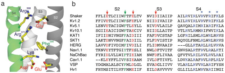
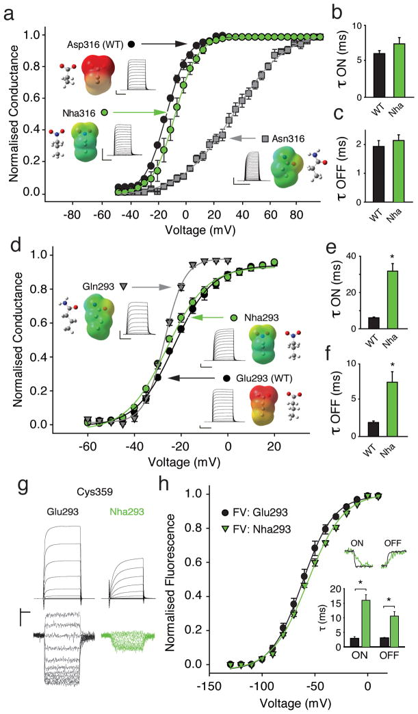
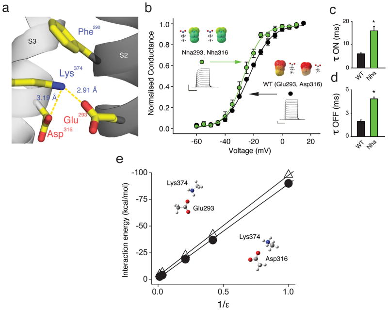
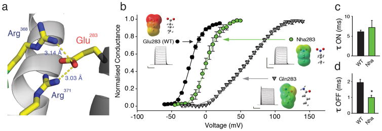
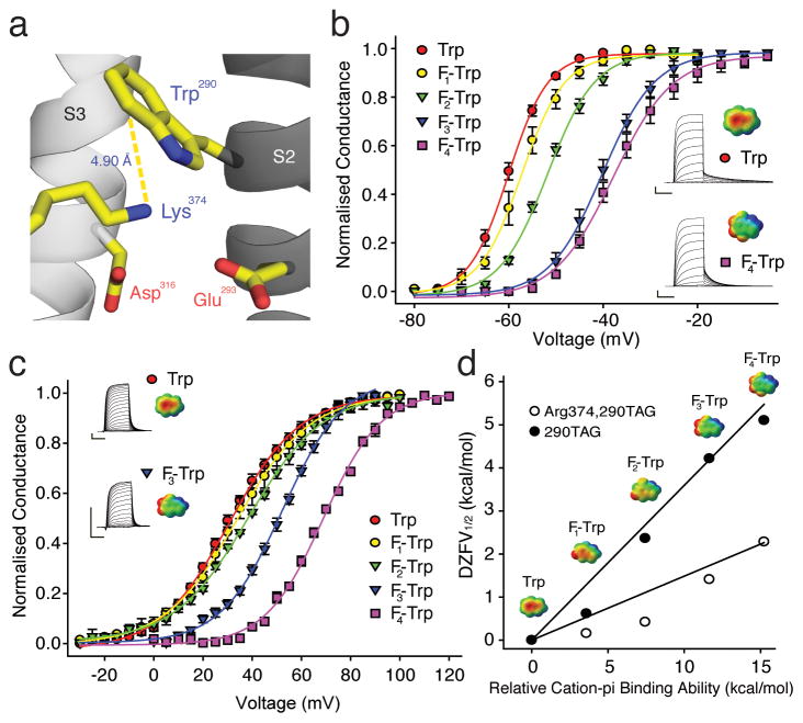
Voltage-sensor domains couple membrane potential to conformational changes in voltage-gated ion channels and phosphatases. Highly coevolved acidic and aromatic side chains assist the transfer of cationic side chains across the transmembrane electric field during voltage sensing. We investigated the functional contribution of negative electrostatic potentials from these residues to channel gating and voltage sensing with unnatural amino acid mutagenesis, electrophysiology, voltage-clamp fluorometry and ab initio calculations. The data show that neutralization of two conserved acidic side chains in transmembrane segments S2 and S3, namely Glu293 and Asp316 in Shaker potassium channels, has little functional effect on conductance-voltage relationships, although Glu293 appears to catalyze S4 movement. Our results suggest that neither Glu293 nor Asp316 engages in electrostatic state-dependent charge-charge interactions with S4, likely because they occupy, and possibly help create, a water-filled vestibule.


Conflict of interest statement

Competing financial interests

The authors declare that they have no competing interests as defined by Nature Publishing Group, or other interests that might be perceived to influence the results and/or discussion reported in this paper.

Figures

Figure 1
Figure 1. Model of transmembrane segments 2–4 and sequence alignment
(a) Model showing transmembrane segments 2 (dark grey), 3 (light grey) and 4 (green), highlighted are Glu283, Glu293, Asp316, Arg 368, Arg371 and Lys374 (PDB 2R9R); (b) Sequence alignment of various voltage-dependent membrane proteins: Shaker (GI: 13432103), Kv1.2 (GI:52000923), Kv5.1 (GI: 20070166), Kv10.1 (GI: 22164088), KAT1 (GI:15237407), SKT1 (GI:1514649), HERG (GI:4557729), Nav1.1 (115583677), Cav1.1 (GI:110349767), NaChBac (GI:10174118), VSP (GI:76253898), Hv1 (GI:91992155). Conserved residues are color-coded: acidic = red, basic = blue and aromatic = green. The arrows indicate residues studied here, the asterisks indicate Arg368, Arg371 and Lys374.
Figure 2
Figure 2. Contributions of Asp 316 and Glu293
(a) GV of natural (Asp, Asn) and unnatural (Nha) side-chains at position 316 (Asn316: V1/2 = 27.8 ± 3.3 mV; Nha316: V1/2 = −17.5 ± 2.3 mV; Asp316 (WT): V1/2 = −22.1 ± 0.7 mV, (n = 5–6)); insets show currents, electrostatic surface potential (ESP, red = −100 kcal/mol, green = 0 kcal/mol, blue = +100 kcal/mol) maps and energy minimized structures of side-chains; (b)/(c) Activation time constants (−80 mV to −25 mV) and deactivation time constants (+20 mV to −25 mV) for Asp316 and Nha316 (single exponential fit, n = 5); (d) GV of natural (Glu, Gln) and unnatural (Nha) side-chains at position 293; insets as in (a); (Gln293: V1/2 = −27.1 ± 0.5 mV; Nha293: V1/2 = −24.8 ± 0.8 mV; Glu293 (WT): V1/2 = −22.1 ± 0.7 mV (n = 4–6)); (e)/(f) Time constants of activation (e) and deactivation (f) for Glu293 and Nha293 (n = 5–7); protocol as in (b)/(c)); (g) Current (upper panel) and fluorescence (lower panel) recordings for Glu293 (WT) or Nha293 background, in black and green, respectively; (h) FV for Glu293 and Nha293 (V1/2: −63.1 ± 3.0 mV and 58.3 ± 1.8 mV, respectively; n = 4). Upper inset shows fluorescence sample traces (ON: −80 mV to −50 mV, OFF: +20 mV to −50 mV); lower inset shows averaged time constants (n = 4). Scale bars: 2 μA for current, 1 % for fluorescence and 50 ms for time (except 20 ms for Asn316). Asterisk indicates significant difference (p < 0.05).
Figure 3
Figure 3. Glu293 and Asp316 do not contribute to a network of electrostatic charge-charge interactions
(a) Model highlighting the proximity of Glu293 and Asp316 to Lys374 (PDB 2R9R); (b) Incorporating Nha simultaneously at positions 293 and 316 has only a minor effect on the GV (V1/2 = −27.2 ± 0.9 mV for Nha at positions 293 and 316 vs. V1/2 = −22.1 ± 0.7 mV for Glu in position 293 and Asp in position 316 (WT), n = 5); insets show currents, ESP maps and energy minimized structures with scale as in Fig. 2a; (c)/(d) Time constants of activation obtained from a depolarizing pulse from −80 mV to −25 mV (c) and time constants of deactivation obtained from a repolarizing pulse from +20 mV to −25 mV (d) for WT and for Nha simultaneously at positions 293 and 316 (all data fit with a single exponential, n = 5); asterisk indicates significant difference (p < 0.05); (e) Plot shows ab initio interaction energies at the HF/6-31G*+ level between Lys374 and Glu293 or Asp316 (open triangles or filled circles, respectively) in different dielectric environments. Side-chain moieties from PDB 2R9R were isolated, fixed and modeled as acetate (Glu and Asp) and methylammonium (Lys) in a variety of dielectric environments. All scale bars: 2 μA for current and 50 ms for time.
Figure 4
Figure 4. Glu283 is likely to form a state-dependent electrostatic charge-charge interaction with S4 charges
(a) Model highlighting the proximity of Glu283 to Arg 368 and Arg371 (PDB 2R9R); (b) Effects on GV by natural (Glu, Gln) and unnatural (Nha) side-chains at position 283 (V1/2 = 58.1 ± 2.0 mV for Gln283 (n = 4); V1/2 = 0.9 ± 2.1 mV for Nha283 (n = 8) and V1/2 = −22.1 ± 0.7 mV for Glu283 (WT)); insets show currents, ESP maps and energy minimized structures of natural and unnatural amino acids used at position 283 with scale for ESPs as in Fig. 2a; (c)/(d) Time constants of activation obtained from a depolarizing pulse from −80 mV to −25 mV (c) and time constants of deactivation obtained from a repolarizing pulse from from +20 mV (WT) or +40 mV (Nha283) to −25 mV (d) with Nha at positions 293 and 316 (all data fit with a single exponential, n = 5); asterisk indicates significant difference (p < 0.05); Scale bars: 2 μA for current, 50 ms for time.
Figure 5
Figure 5. A cation-pi interaction in the potassium channel voltage-sensor
(a) A model highlighting the proximity of Trp290 and Lys374 (PDB 3LNM); (b) Effect of fluorination at Trp290 on GV; insets show currents and ESPs for Trp (upper) and 4,5,6,7-F4-Trp (lower) (n = 5–7); scale for ESPs: red = −25 kcal/mol, green = 0 kcal/mol, blue = +25 kcal/mol; Scale bars: 2 μA for current, 50 ms for time; (c) Effect of fluorination at Trp290 on the Arg374 background; insets show currents and ESPs for Trp (upper) and 5,6,7-F4-Trp (lower) (n = 4–6); scale for ESPs as in (b); (d) Cation-pi plot for fluorination of Trp290 with Lys or Arg in position 374. ESPs for Trp derivatives are shown with scales as in (b).

References

    1. Hille B. Ion channels of excitable membranes. 3. Sinauer; 2001.
    1. Murata Y, Iwasaki H, Sasaki M, Inaba K, Okamura Y. Phosphoinositide phosphatase activity coupled to an intrinsic voltage sensor. Nature. 2005;435:1239–1243. - PubMed
    1. Ramsey IS, Moran MM, Chong JA, Clapham DE. A voltage-gated proton-selective channel lacking the pore domain. Nature. 2006;440:1213–1216. - PMC - PubMed
    1. Ahern CA, Horn R. Focused electric field across the voltage sensor of potassium channels. Neuron. 2005;48:25–29. - PubMed
    1. Aggarwal SK, MacKinnon R. Contribution of the S4 segment to gating charge in the Shaker K+ channel. Neuron. 1996;16:1169–1177. - PubMed

Publication types