. 2011:2011:569416.
doi: 10.4061/2011/569416. Epub 2011 Jul 12.

Cytotoxicity and induction of inflammation by pepsin in Acid in bronchial epithelial cells

Affiliations

Cytotoxicity and induction of inflammation by pepsin in Acid in bronchial epithelial cells

Erik Bathoorn et al. Int J Inflam. 2011.

Abstract

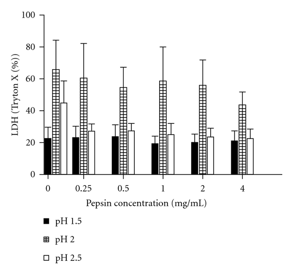
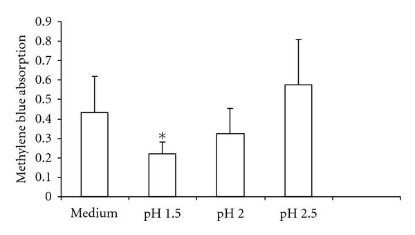
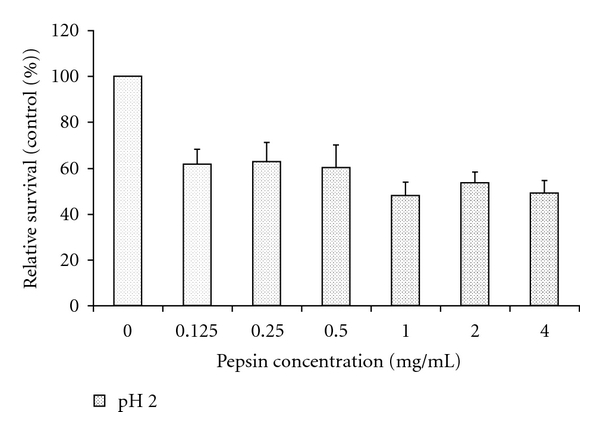
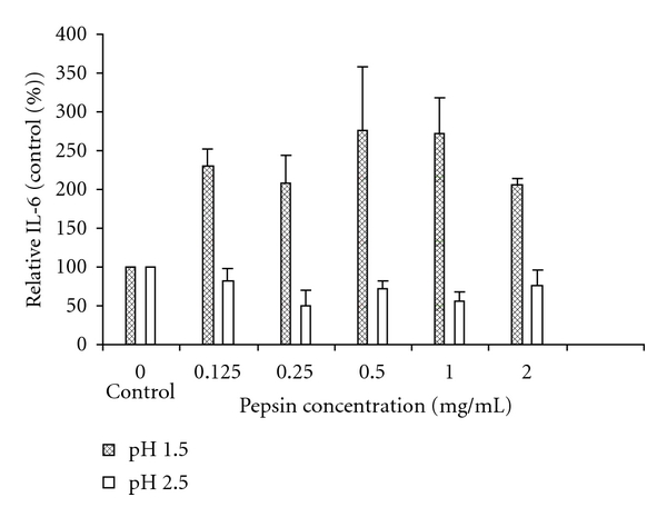
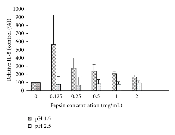
Introduction. Gastroesophageal reflux has been associated with chronic inflammatory diseases and may be a cause of airway remodelling. Aspiration of gastric fluids may cause damage to airway epithelial cells, not only because acidity is toxic to bronchial epithelial cells, but also since it contains digestive enzymes, such as pepsin. Aim. To study whether pepsin enhances cytotoxicity and inflammation in airway epithelial cells, and whether this is pH-dependent. Methods. Human bronchial epithelial cells were exposed to increasing pepsin concentrations in varying acidic milieus, and cell proliferation and cytokine release were assessed. Results. Cell survival was decreased by pepsin exposure depending on its concentration (F = 17.4) and pH level of the medium (F = 6.5) (both P < 0.01). Pepsin-induced interleukin-8 release was greater at lower pH (F = 5.1; P < 0.01). Interleukin-6 induction by pepsin was greater at pH 1.5 compared to pH 2.5 (mean difference 434%; P = 0.03). Conclusion. Pepsin is cytotoxic to bronchial epithelial cells and induces inflammation in addition to acid alone, dependent on the level of acidity. Future studies should assess whether chronic aspiration causes airway remodelling in chronic inflammatory lung diseases.


Figures

Figure 1
Figure 1
Exposure of cells for 5 minutes induced more cytotoxicity as measured by LDH after 20 hours incubation at pH 2.0 (mean (SEM) 57%  ± 5.77%) compared to both pH 1.5 and pH 2.5 (22%  ± 2.17%, and 28%  ± 2.81%, resp., both P < 0.01). LDH release was pH-dependent (P < 0.01). There was a trend for a pepsin effect (P = 0.078), with lower LDH release with pepsin at all concentrations compared to no pepsin, independent of pH levels.
Figure 2
Figure 2
pH dependence of 16-HBE cell survival, as measured by methylene blue absorption. *Exposure of cells to medium with pH 1.5 for 5 minutes resulted in significantly less cell proliferation after 20 hours of incubation compared to exposure to pH 2.5 (mean absorption 0.22 versus 0.57; P = 0.03, n = 3). pH of control medium was 7.9. Histograms represent the means and the bars the SEM.
Figure 3
Figure 3
Effects of pepsin on cell survival, as measured by methylene blue absorption, and expressed as a % of absorbance of treatment with the corresponding pH without pepsin (control). Pepsin induced a significant decrease in cell survival independent of pH (F = 6.5; P < 0.01). Data are expressed as means (histograms) and SEM (bars) for 3 replicates. See supplement Figure  1 for the effect of pepsin on cell survival of all the pH levels used in the statistical analysis.
Figure 4
Figure 4
Interleukin-6 production by 16-HBE cells, corrected for cell proliferation and expressed as a % of exposure with the corresponding pH without pepsin (control). Interleukin-6 release induced by pepsin is higher at pH 1.5 compared to pH 2.5 (mean difference 283%; P = 0.03). Data are expressed as means (histograms) and SEM (bars) for 3 replicates. See supplement Figure  2 for induction of IL-6 at all pepsin concentrations and pH levels used in the statistical analysis.
Figure 5
Figure 5
Interleukin-8 production by 16-HBE cells, corrected for cell proliferation and expressed as a % of exposure with the corresponding pH without pepsin (control). Interleukin-8 release induced by pepsin is higher at pH 1.5 compared to pH 2.5 (mean difference 221%; P < 0.01). Data expressed as mean (histograms) and SEM (bars) for 3 replicates. See supplement Figure  3 for induction of IL-8 at all pepsin concentrations and pH levels used in the statistical analysis.

References

    1. Wynne JW, Ramphal R, Hood CI. Tracheal mucosal damage after aspiration. A scanning electron microscope study. American Review of Respiratory Disease. 1981;124(6):728–732. - PubMed
    1. Bonnans C, Fukunaga K, Levy MA, Levy BD. Lipoxin A4 regulates bronchial epithelial cell responses to acid injury. American Journal of Pathology. 2006;168(4):1064–1072. - PMC - PubMed
    1. Beck-Schimmer B, Rosenberger DS, Neff SB, et al. Pulmonary aspiration: new therapeutic approaches in the experimental model. Anesthesiology. 2005;103(3):556–566. - PubMed
    1. Nader ND, Davidson BA, Tait AR, Holm BA, Knight PR. Serine antiproteinase administration preserves innate superoxide dismutase levels after acid aspiration and hyperoxia but does not decrease lung injury. Anesthesia and Analgesia. 2005;101(1):213–219. - PubMed
    1. Nagase T, Uozumi N, Ishii S, et al. Acute lung injury by sepsis and acid aspiration: a key role for cytosolic phospholipase A2. Nature Immunology. 2000;1(1):42–45. - PubMed

LinkOut - more resources