

Evaluation of computer-assisted quantification of carotid artery stenosis
- PMID: 21786073
- PMCID: PMC3295974
- DOI: 10.1007/s10278-011-9413-y
Evaluation of computer-assisted quantification of carotid artery stenosis
Abstract
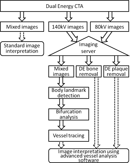
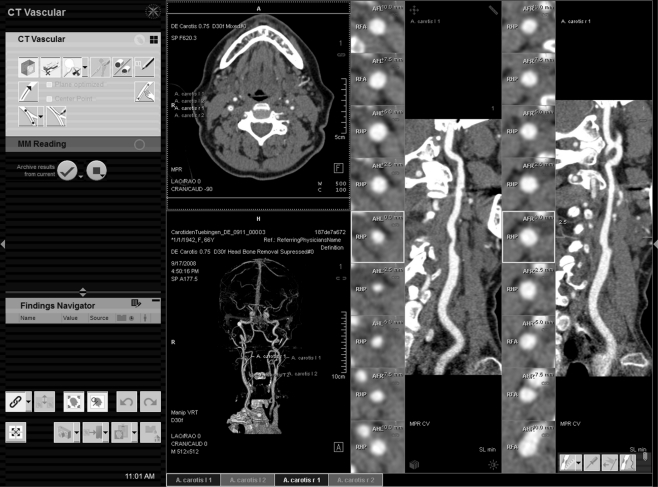
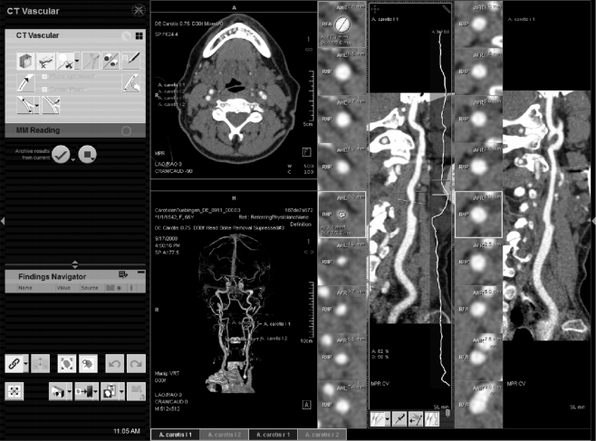
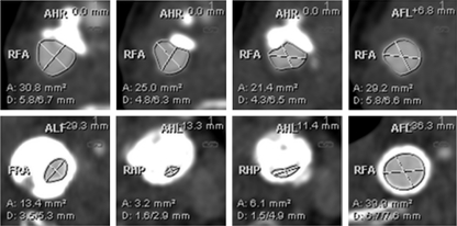
The purpose of this study was to evaluate the influence of advanced software assistance on the assessment of carotid artery stenosis; particularly, the inter-observer variability of readers with different level of experience is to be investigated. Forty patients with suspected carotid artery stenosis received head and neck dual-energy CT angiography as part of their pre-interventional workup. Four blinded readers with different levels of experience performed standard imaging interpretation. At least 1 day later, they performed quantification using an advanced vessel analysis software including automatic dual-energy bone and hard plaque removal, automatic and semiautomatic vessel segmentation, as well as creation of curved planar reformation. Results were evaluated for the reproducibility of stenosis quantification of different readers by calculating the kappa and correlation values. Consensus reading of the two most experienced readers was used as the standard of reference. For standard imaging interpretation, experienced readers reached very good (k = 0.85) and good (k = 0.78) inter-observer variability. Inexperienced readers achieved moderate (k = 0.6) and fair (k = 0.24) results. Sensitivity values 80%, 91%, 83%, 77% and specificity values 100%, 84%, 82%, 53% were achieved for significant area stenosis >70%. For grading using advanced vessel analysis software, all readers achieved good inter-observer variability (k = 0.77, 0.72, 0.71, and 0.77). Specificity values of 97%, 95%, 95%, 93% and sensitivity values of 84%, 78%, 86%, 92% were achieved. In conclusion, when supported by advanced vessel analysis software, experienced readers are able to achieve good reproducibility. Even inexperienced readers are able to achieve good results in the assessment of carotid artery stenosis when using advanced vessel analysis software.
Figures
References
-
- Hollingworth W, Nathens AB, Kanne JP, Crandall ML, Crummy TA, Hallam DK, Wang MC, Jarvik JG. The diagnostic accuracy of computed tomography angiography for traumatic or atherosclerotic lesions of the carotid and vertebral arteries: a systematic review. Eur J Radiol. 2003;48:88–102. doi: 10.1016/S0720-048X(03)00200-6. - DOI - PubMed
-
- Josephson SA, Bryant SO, Mak HK, Johnston SC, Dillon WP, Smith WS. Evaluation of carotid stenosis using ct angiography in the initial evaluation of stroke and TIA. Neurology. 2004;63:457–460. - PubMed
-
- Koelemay MJW, Nederkoorn PJ, Reitsma JB, Majoie CB. Systematic review of computed tomographic angiography for assessment of carotid artery disease. Stroke. 2004;35:2306–2312. doi: 10.1161/01.STR.0000141426.63959.cc. - DOI - PubMed
MeSH terms
Substances
LinkOut - more resources
Full Text Sources
Other Literature Sources
Medical
