

Progressive multifocal leukoencephalopathy associated with isolated CD8+ T-lymphocyte deficiency mimicking tumefactive MS
- PMID: 21786075
- PMCID: PMC3204182
- DOI: 10.1007/s13365-011-0045-2
Progressive multifocal leukoencephalopathy associated with isolated CD8+ T-lymphocyte deficiency mimicking tumefactive MS
Abstract
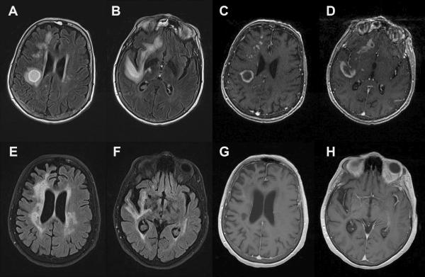
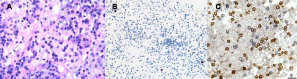
Progressive multifocal leukoencephalopathy (PML) is a severe demyelinating disease of the central nervous system (CNS) caused by lytic infection of oligodendrocytes by the polyomavirus JC (JCV). PML has classically been described in individuals with profound cellular immunosuppression. While some case reports have documented PML in individuals with minimal or occult immunosuppression, such cases are very rare and their pathogenesis is not well understood. We report a unique case of a 74 year-old woman who developed PML clinically mimicking tumefactive multiple sclerosis in the context of an idiopathic isolated CD8+ T-lymphocytopenia. Her course subsequently stabilized, concomitant to the development of a cellular immune response directed against JCV. We review the current literature of related cases and discuss the pathogenesis and implications of this rare presentation.
Figures
Similar articles
-
Brain Biopsy Is More Reliable than the DNA test for JC Virus in Cerebrospinal Fluid for the Diagnosis of Progressive Multifocal Leukoencephalopathy.Intern Med. 2017;56(10):1231-1234. doi: 10.2169/internalmedicine.56.7689. Epub 2017 May 15. Intern Med. 2017. PMID: 28502942 Free PMC article.
-
Progressive multifocal leukoencephalopathy in a patient with pre-clinical primary biliary cirrhosis.Clin Neurol Neurosurg. 2014 Aug;123:45-9. doi: 10.1016/j.clineuro.2014.04.032. Epub 2014 May 10. Clin Neurol Neurosurg. 2014. PMID: 25012010 No abstract available.
-
Progressive multifocal leukoencephalopathy in common variable immunodeficiency: mitigated course under mirtazapine and mefloquine.J Neurovirol. 2015 Dec;21(6):694-701. doi: 10.1007/s13365-015-0340-4. Epub 2015 Apr 28. J Neurovirol. 2015. PMID: 25916731
-
[Idiopathic CD4-positive lymphocytopenia-associated progressive multifocal leukoencephalopathy confirmed by brain biopsy following negative results of repeated CSF-JC-virus tests: a case report].Rinsho Shinkeigaku. 2018 Dec 21;58(12):750-755. doi: 10.5692/clinicalneurol.cn-001227. Epub 2018 Nov 29. Rinsho Shinkeigaku. 2018. PMID: 30487366 Review. Japanese.
-
Asymptomatic progressive multifocal leukoencephalopathy: a case report and review of the literature.J Med Case Rep. 2018 Jul 1;12(1):187. doi: 10.1186/s13256-018-1727-7. J Med Case Rep. 2018. PMID: 29960601 Free PMC article. Review.
Cited by
-
The Use of Antimalarial Drugs against Viral Infection.Microorganisms. 2020 Jan 8;8(1):85. doi: 10.3390/microorganisms8010085. Microorganisms. 2020. PMID: 31936284 Free PMC article. Review.
-
Progressive multifocal leukoencephalopathy successfully treated with mefloquine and literature review.Encephalitis. 2021 Oct;1(4):111-119. doi: 10.47936/encephalitis.2021.00094. Epub 2021 Sep 28. Encephalitis. 2021. PMID: 37470049 Free PMC article.
-
Progressive neurologic dysfunction in a psoriasis patient treated with dimethyl fumarate.Ann Neurol. 2015 Oct;78(4):501-14. doi: 10.1002/ana.24471. Epub 2015 Jul 31. Ann Neurol. 2015. PMID: 26150206 Free PMC article. Review.
-
Progressive Multifocal Leukoencephalopathy Associated With Idiopathic CD8+ Lymphocytopenia.Cureus. 2022 Dec 23;14(12):e32870. doi: 10.7759/cureus.32870. eCollection 2022 Dec. Cureus. 2022. PMID: 36694528 Free PMC article.
References
-
- Berger JR, Levy RM, Flomenhoft D, Dobbs M. Predictive factors for prolonged survival in acquired immunodeficiency syndrome-associated progressive multifocal leukoencephalopathy. Ann Neurol. 1998;44(3):341–349. - PubMed
-
- Berger JR, Pall L, Lanska D, Whiteman M. Progressive multifocal leukoencephalopathy in patients with HIV infection. J Neurovirol. 1998;4(1):59–68. - PubMed
-
- Boerman R, Bax J, Beekhuis-Brussee J, Medaer R, Bollen L. JC virus and multiple sclerosis: a refutation? Acta Neurol Scand. 1993;87:353–355. - PubMed
Publication types
MeSH terms
Substances
Grants and funding
LinkOut - more resources
Full Text Sources
Medical
Research Materials
