Review
. 2011 Dec;68(24):3995-4008.
doi: 10.1007/s00018-011-0770-y. Epub 2011 Jul 24.

Specification of neuronal and glial subtypes from human pluripotent stem cells

Affiliations
Review

Specification of neuronal and glial subtypes from human pluripotent stem cells

Huisheng Liu et al. Cell Mol Life Sci. 2011 Dec.

Abstract

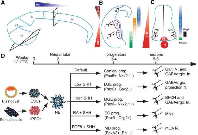
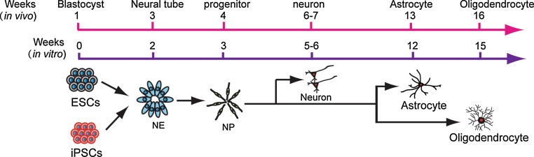
Human pluripotent stem cells (PSCs), including embryonic stem cells (ESCs) and induced pluripotent stem cells (iPSCs), provide a dynamic tool for revealing early embryonic development, modeling pathological processes, and developing therapeutics through drug discovery and potential cell replacement. The first step toward the utilities of human PSCs is directed differentiation to functionally specialized cell/tissue types. Following developmental principles, human ESCs, and lately iPSCs, have been effectively differentiated to region- and/or transmitter-specific neuronal and glial types, including cerebral glutamatergic, striatal γ-aminobutyric acid (GABA)-ergic, forebrain cholinergic, midbrain dopaminergic, and spinal motor neurons, as well as astrocytes and oligodendrocytes. These studies also reveal unique aspects of human cell biology, including intrinsically programmed developmental course, differential uses of transcription factors for neuroectoderm specification, and distinct responses to extracellular signals in regulating cell fate. Such information will be instrumental in translating biological findings to therapeutic development.


Figures

Fig. 1
Fig. 1
Neuronal subtype specification in vivo and vitro. A The developing brain is patterned by morphogens like FGFs and RA along the anterior-posterior axis into forebrain (FB), middle brain (MB), hindbrain (HB), and spinal cord (SC). In each part of the brain and spinal cord, it is further subdivided into multiple domains along the dorsal–ventral axis by morphogens like WNTs, BMPs, and SHH. B In the forebrain, as shown in coronal hemi-section through 1 in a, two opposing morphogens, ventrally derived SHH and dorsally originated WNTs define the cortical, LGE and MGE domains. Progenitors in these domains mainly generate glutamatergic neurons (a), GABAergic projection neurons (b), and BFCNs (c), respectively. GABAergic interneurons are differentiated from all three domains in human. C Similarly, in the spinal cord, as shown in the cross section from site 2 in A, SHH gradients released from notochord (N) and floor plate (FP) define ventral domains in the ventral spinal cord and MNs are generated from the Olig2-expressing ventral progenitors (prog.). D In parallel with in vivo neural development, human ESCs and iPSCs are first differentiated toward Pax6-expressing primitive neuroepithelia (NE) in the absence of morphogens in the first week. In the absence of exogenous morphogens, the NE differentiate to Pax6-expressing cortical progenitors, and subsequently generate glutamatergic neurons (Glut. N.) and GABAergic interneurons (GABAergic In.) by default. In the presence of a low concentration of SHH, the NE become Gsx-expressing LGE progenitors and they later generate DARPP32-expressing GABAergic projection neurons. With higher SHH concentration, the NE are fated to MGE progenitors, which produce BFCNs and GABAergic interneurons. Under RA, the primitive NE are caudalized to hindbrain/spinal cord progenitors, which become Olig2-expressing progenitors in the presence of SHH and then differentiate to motor neurons (MNs). In the presence of FGF8 and SHH, the primitive NE are specified to ventral midbrain progenitors, which then produce mDA neurons
Fig. 2
Fig. 2
Temporal course of glial differentiation in vivo and in vitro. In parallel with in vivo development, human PSCs begin to generate neurons in the second month, astrocytes in the third month, and oligodendrocytes in the fourth month

References

    1. Thomson JA, Itskovitz-Eldor J, Shapiro SS, Waknitz MA, Swiergiel JJ, Marshall VS, Jones JM. Embryonic stem cell lines derived from human blastocysts. Science. 1998;282:1145–1147. doi: 10.1126/science.282.5391.1145. - DOI - PubMed
    1. Yu J, Vodyanik MA, Smuga-Otto K, Antosiewicz-Bourget J, Frane JL, Tian S, Nie J, Jonsdottir GA, Ruotti V, Stewart R, Slukvin Ii, Thomson JA. Induced pluripotent stem cell lines derived from human somatic cells. Science. 2007;318:1917–1920. doi: 10.1126/science.1151526. - DOI - PubMed
    1. Takahashi K, Yamanaka S. Induction of pluripotent stem cells from mouse embryonic and adult fibroblast cultures by defined factors. Cell. 2006;126:663–676. doi: 10.1016/j.cell.2006.07.024. - DOI - PubMed
    1. Takahashi K, Tanabe K, Ohnuki M, Narita M, Ichisaka T, Tomoda K, Yamanaka S. Induction of pluripotent stem cells from adult human fibroblasts by defined factors. Cell. 2007;131:861–872. doi: 10.1016/j.cell.2007.11.019. - DOI - PubMed
    1. Pang ZP, Yang N, Vierbuchen T, Ostermeier A, Fuentes DR, Yang TQ, Citri A, Sebastiano V, Marro S, Sudhof TC, Wernig M (2011) Induction of human neuronal cells by defined transcription factors. Nature [Epub ahead of print] - PMC - PubMed

Publication types

MeSH terms

LinkOut - more resources