. 2011 Oct;29(10):1537-48.
doi: 10.1002/stem.697.

Mesenchymal stem cells expressing insulin-like growth factor-I (MSCIGF) promote fracture healing and restore new bone formation in Irs1 knockout mice: analyses of MSCIGF autocrine and paracrine regenerative effects

Affiliations

Mesenchymal stem cells expressing insulin-like growth factor-I (MSCIGF) promote fracture healing and restore new bone formation in Irs1 knockout mice: analyses of MSCIGF autocrine and paracrine regenerative effects

Froilán Granero-Moltó et al. Stem Cells. 2011 Oct.

Abstract

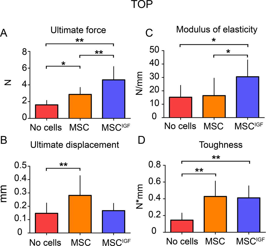
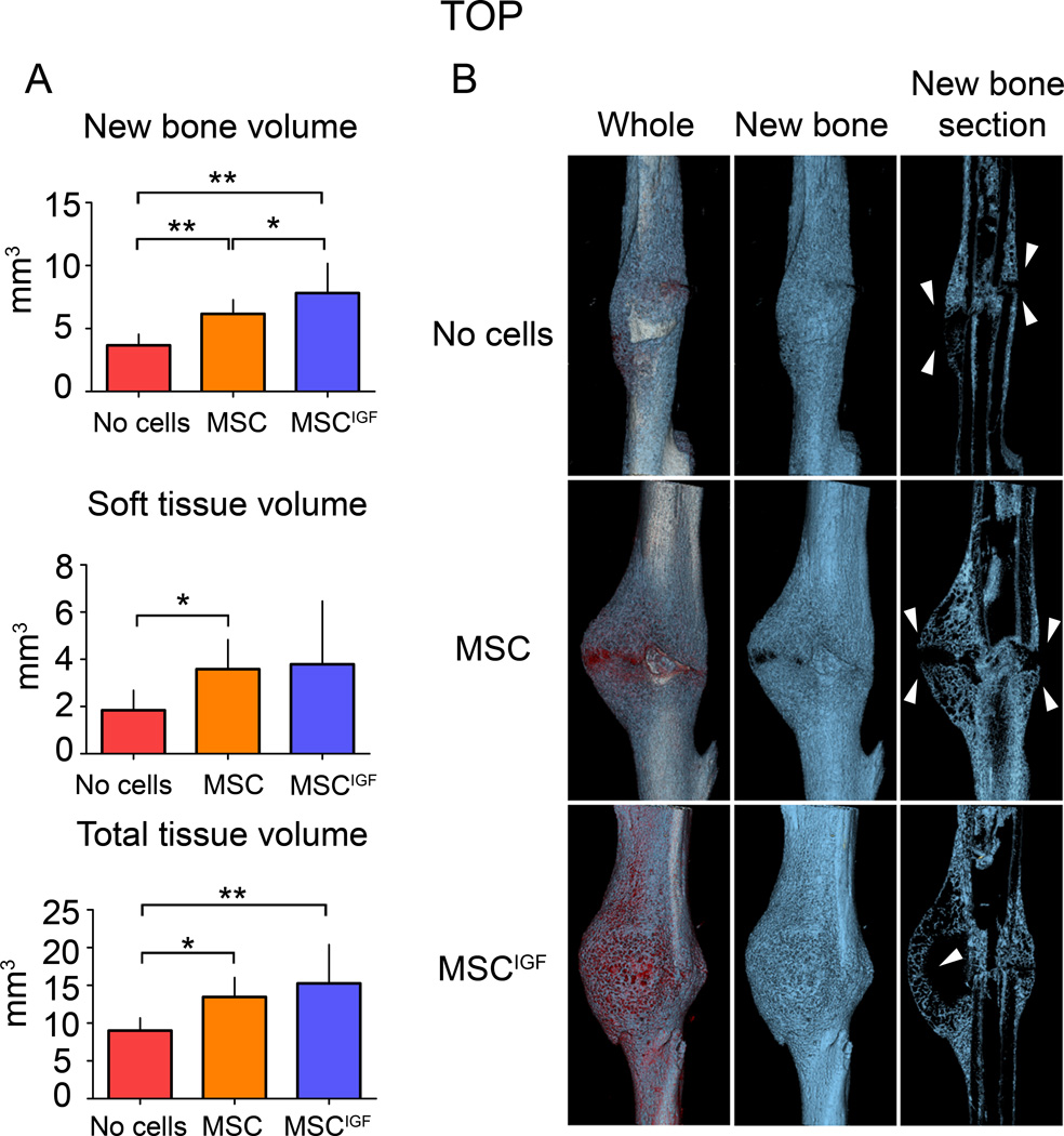
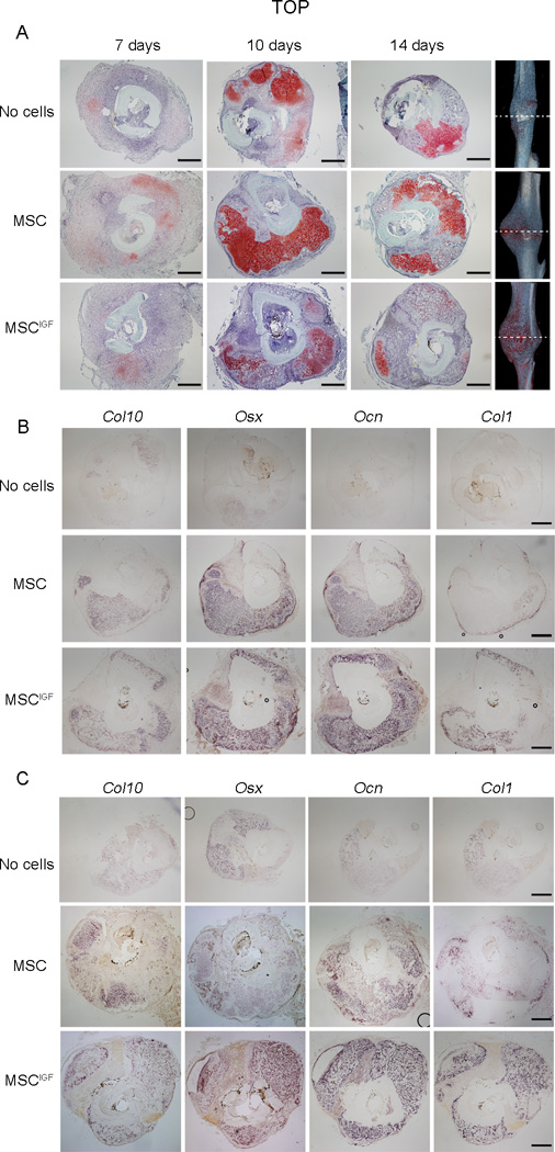
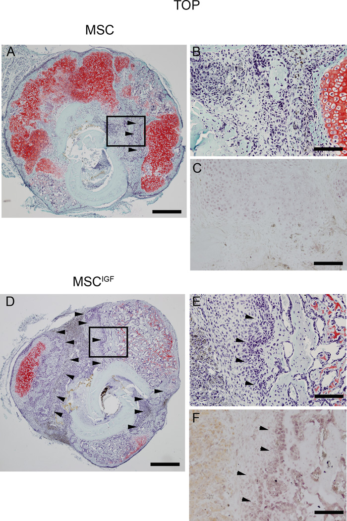
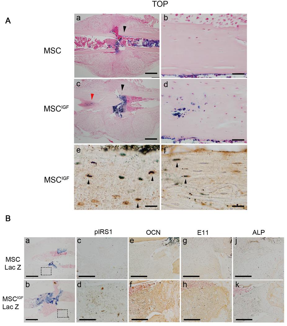
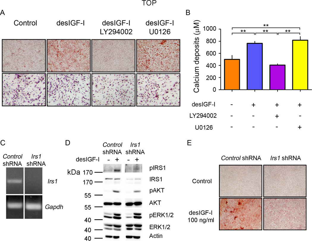
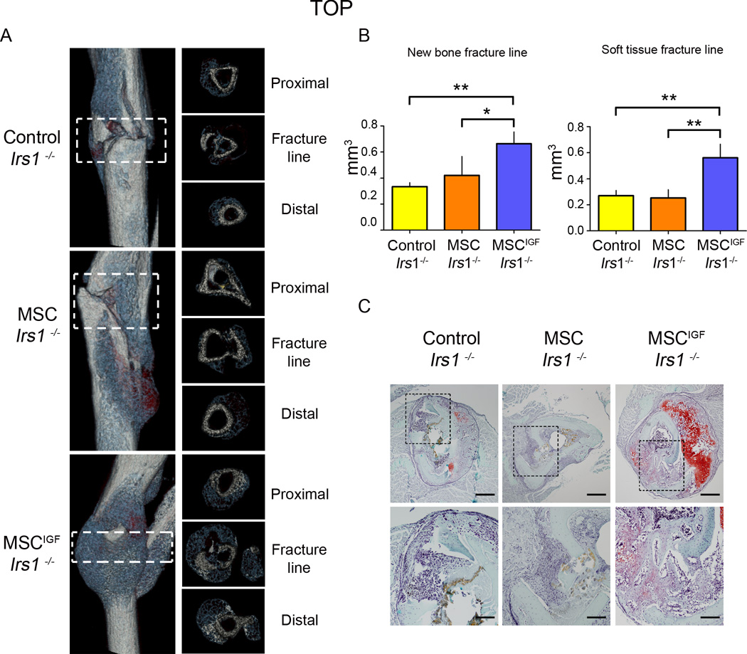
Failures of fracture repair (nonunions) occur in 10% of all fractures. The use of mesenchymal stem cells (MSC) in tissue regeneration appears to be rationale, safe, and feasible. The contributions of MSC to the reparative process can occur through autocrine and paracrine effects. The primary objective of this study is to find a novel mean, by transplanting primary cultures of bone marrow-derived MSCs expressing insulin-like growth factor-I (MSC(IGF)), to promote these seed-and-soil actions of MSC to fully implement their regenerative abilities in fracture repair and nonunions. MSC(IGF) or traceable MSC(IGF)-Lac-Z were transplanted into wild-type or insulin-receptor-substrate knockout (Irs1(-/-)) mice with a stabilized tibia fracture. Healing was assessed using biomechanical testing, microcomputed tomography (μCT), and histological analyses. We found that systemically transplanted MSC(IGF) through autocrine and paracrine actions improved the fracture mechanical strength and increased new bone content while accelerating mineralization. We determined that IGF-I adapted the response of transplanted MSC(IGF) to promote their differentiation into osteoblasts. In vitro and in vivo studies showed that IGF-I-induced osteoglastogenesis in MSCs was dependent of an intact IRS1-PI3K signaling. Furthermore, using Irs1(-/-) mice as a nonunion fracture model through altered IGF signaling, we demonstrated that the autocrine effect of IGF-I on MSC restored the fracture new bone formation and promoted the occurrence of a well-organized callus that bridged the gap. A callus that was basically absent in Irs1(-/-) left untransplanted or transplanted with MSCs. We provided evidence of effects and mechanisms for transplanted MSC(IGF) in fracture repair and potentially to treat nonunions.


Conflict of interest statement

Disclosure of potential conflict of interest.

The authors indicate no potential conflicts of interest.

Figures

Figure 1
Figure 1
Transplanted MSCIGF improve the callus biomechanical properties differently from MSC. Fourteen days after tibia fracture, calluses from mice that had received either MSC or MSCIGF transplant after the fracture or remained untransplanted (No cells) were dissected and subjected to distraction to failure BMT. p values of one-way ANOVA analyses are respectively: p=0.0001 for ultimate force; p=0.02 for ultimate displacement; p=0.02 for modulus of elasticity; p=0.0002 for toughness. *, p<0.05, **, p<0.01 by Bonferroni post hoc test. No cells, n=11, MSC, n=10, MSCIGF, n=5.
Figure 2
Figure 2
MSCIGF transplant increases the fracture callus new bone content. µCT analyses were performed on 14 day post-fracture tibias in transplanted mice (either with MSC or MSCIGF) and untransplanted mice (No cells). A, total tissue volume, new bone volume (blue) and soft tissue volume (red) were calculated as reported in the Supplemental Methods section. New bone volume, ANOVA p-value=0.0006; soft tissue, ANOVA p-value=0.0395; total tissue volume, ANOVA p-value=0.0058. *, p<0.05; **, p<0.01 by Bonferroni post hoc test.. No cells, n=7; MSC, n=10; MSCIGF, n=7. B, Three dimensional reconstruction of representative calluses from control (No cells) and MSC transplanted mice (MSC, MSCIGF).
Figure 3
Figure 3
MSCIGF transplant induces bone formation that bridges the fracture gap. Histological sections were obtained from calluses of either MSC or MSCIGF recipients or from mice that were left in transplanted (No cells) respectively 7, 10 and 14 days after fracture. A, Serial cross-sections of 7, 10 and 14 days fracture calluses were visualized with Safranin O/Fast Green staining for the presence of cartilage (red) and bone (green), depicted are sections of the fracture line. Ten (B) and fourteen (C) days post fracture calluses were analyzed by in situ hybridization for the presence of bone and cartilage markers namely Col10, Osx, Ocn and Col1. Bar, 500 µm.
Figure 4
Figure 4
MSCIGF transplant increases the areas of intramembraneous ossification. Serial cross-sections of 14 days post-fracture calluses from mice either transplanted with MSC (A,B,C) or MSCIGF (D,E,F) were subjected to Safranin O/Fast Green staining to visualize the presence of cartilage (red) and bone (green). In MSCIGF recipients the Fast Green stainable areas of intramembraneous ossification were also positive for Osx detected by in situ hybridization (C, F) (arrowheads). Bar, 500 µm (A,D). 100 µm (B,C,E,F).
Figure 5
Figure 5
IGF-I induces MSC osteoblastic differentiation in vivo through autocrine and paracrine mechanisms. Mice were transplanted with either MSC-Lac-Z or MSCIGF–Lac-Z. Calluses were obtained 7 days after fracture and subjected to Lac-Z staining. A, (a), MSC-Lac-Z positive cells localize within the calluses at the endosteum, bone marrow and fracture rim. (c), in addition to these locations, MSCIGF-Lac-Z positive cells localize in the cortical bone (arrow heads). (d), magnification of the area indicated by the black arrow head in (c). (e), histological sections from MSCIGF–Lac-Z recipients adjacent to the section depicted in (a), were subjected to IHC for DMP-1 following Lac-Z staining. DMP-1-Lac-Z double positive cells are depicted as dark brown cells (indicated by arrow-heads); DMP1 single positive in orange; Lac-Z single positive (none in this section) as light blue. (f), histological sections from MSCIGF–Lac-Z recipients adjacent to the section depicted in (a), were subjected to IHC for OCN following Lac-Z staining. OCN-Lac-Z double positive cells are depicted as dark brown cells (indicated by arrow-heads); OCN single positive in orange; Lac-Z single positive (none in this section) as light blue. B, calluses from mice that received either MSC-Lac Z or MSCIGF-Lac Z transplant were analyzed at 7 days post-fracture by Lac-Z staining (a and b) and adjacent sections by Lac-Z staining followed by IHC for either pIRS1 (c and d) or OCN (e and f) or E11 (h and h) and ALP (j and k). Dotted squares in (a) indicate areas magnified respectively in (c), (e), (g) and (j); dotted squares in (b) the areas magnified in (d), (f), (h) and (k). Double positive (Lac-Z and IHC) cells are depicted as dark brown, IHC single positive in orange; Lac-Z single positive as light blue. See comments in the text. Bar, A, 500 µm (a, c); 100 µm (b,d); 50 µm(e, f). B, 100 µm (a, b), 50 µm (c,d,e,f,g,h,j,k).
Figure 6
Figure 6
IGF-I induces osteoblastic differentiation of MSC through the PI3K-AKT pathway. A, MSC cultures were grown to confluence and differentiated to osteoblasts in the presence or absence of either LY294002 (10 µM) or U0126 (5 µM) with or without desIGF-I (100 ng/ml) for 14 days. The presence of mineralization was detected by Alizarin Red staining, upper panels. The presence of attached cells was determined by Hematoxylin staining, lower panels. B, Calcium deposits were quantified in MSC cultured in osteogenic medium for 14 days. ANOVA p value=0.0001. **, p<0.01 by Bonferroni ad hoc test. n=3 for each group. C, MSC infected with retroviruses containing a scramble sequence (Control shRNA) or Irs1 specific sequence (Irs1 shRNA) were tested for the presence of Irs1 mRNA by RT-PCR. D, MSC infected with either with control shRNA or Irs1 shRNA were treated with desIGF-I (100 ng/ml) for 10 minutes and protein extracts (10 µg) assayed for the presence of the indicated proteins or phosphorylated specific isoforms (p). E, Control and Irs1 silenced MSC cultures were induced to osteoblast differentiation in osteogenic medium with or without desIGF-I for 14 days (see Methods for more details). The presence of mineralization was detected by Alizarin Red staining.
Figure 7
Figure 7
MSCIGF transplant improves the healing in a model of non-union tibia fracture (Irs1−/−). A, Fourteen days after fracture, dissected tibias of Irs1−/− untransplanted (Control) or transplanted either with MSC or MSCIGF were analyzed by µCT. Depicted are representative three dimensional reconstructions of the calluses from each group (left panel) and cross-sectional sections (right panel). B, New bone and soft tissue content at the fracture line were quantified using a µCT-histological-in situ based thresholding analysis (see Supplemental Methods). The fracture line was defined as the region comprised between the proximal to distal fracture borders, indicated by the dotted squares. Control; Irs1−/−, n=3; MSC Irs1−/−, n=4; MSCIGF Irs−/−, n=3. p values of one-way ANOVA analyses are respectively: new bone, p=0.0178; soft tissue, p=0.0018. *, p<0.05; **, p<0.01 by Bonferroni ad hoc test. C, Sequential cross-sections were stained for the presence of cartilage and bone tissue with Safranin O/Fast Green. Bar, 500 µm (upper panels) , 200 µm (lower panels).

References

    1. Praemer AF, S, Rice DP. Musculoskeletal Conditions in the United States Rosemont, Illinois. 1999.
    1. Axelrad TW, Kakar S, Einhorn TA. New technologies for the enhancement of skeletal repair. Injury. 2007;38(Suppl 1):S49–S62. - PubMed
    1. da Silva Meirelles L, Caplan AI, Nardi NB. In search of the in vivo identity of mesenchymal stem cells. Stem cells (Dayton, Ohio) 2008;26:2287–2299. - PubMed
    1. Nauta AJ, Fibbe WE. Immunomodulatory properties of mesenchymal stromal cells. Blood. 2007;110:3499–3506. - PubMed
    1. Caplan AI. Adult mesenchymal stem cells for tissue engineering versus regenerative medicine. Journal of cellular physiology. 2007;213:341–347. - PubMed

Publication types

MeSH terms

Substances