. 2011 Aug 25;115(33):9948-54.
doi: 10.1021/jp205663z. Epub 2011 Aug 2.

Solid-state NMR spectroscopy provides atomic-level insights into the dehydration of cartilage

Affiliations

Solid-state NMR spectroscopy provides atomic-level insights into the dehydration of cartilage

Jiadi Xu et al. J Phys Chem B. .

Abstract

An atomic-level insight into the functioning of articular cartilage would be useful to develop prevention strategies and therapies for joint diseases such as osteoarthritis. However, the composition and structure of cartilage and their relationship to its unique mechanical properties are quite complex and pose tremendous challenges to most biophysical techniques. In this study, we present an investigation of the structure and dynamics of polymeric molecules of articular cartilage using time-resolved solid-state NMR spectroscopy during dehydration. Full-thickness cartilage explants were used in magic-angle spinning experiments to monitor the structural changes of rigid and mobile carbons. Our results reveal that the dehydration reduced the mobility of collagen amino acid residues and carbon sugar ring structures in glycosaminoglycans but had no effect on the trans-Xaa-Pro conformation. Equally interestingly, our results demonstrate that the dehydration effects are reversible, and the molecular structure and mobility are restored upon rehydration.


Figures

Figure 1
Figure 1
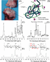
A bovine articular cartilage (A) and its molecular-level representation (B). 13C NMR spectra of cartilage obtained using Ramp-CP (C) and RINEPT (D) pulse sequences under 10 kHz MAS at 25 °C. Spectra were obtained using a Varian VNMRS 600 MHz solid-state NMR spectrometer and a 4 mm double resonance MAS probe. Other experimental parameters include a 2 ms ramp-cross-polarization time, a 80 kHz TPPM proton decoupling during acquisition, and a 3 s recycle delay were used. To obtain the spectrum (D), the RF-free time delays (i.e., all the tau periods in the –tau-180°-tau- sequence of RINEPT) to evolve the transverse magnetization under heteronuclear couplings in RINEPT were set to 500 ms delay to suppress resonances arising from rigid part of the molecules in cartilage. 13C Ramp-CP NMR spectra of hyaluronan (HA) (E) and chondroitin-6-sulfate (CS) (F) powder specimens obtained under 10 kHz MAS.
Figure 2
Figure 2
(A) 1H NMR spectra of cartilage recorded at different times of dehydration and rehydration at PBS buffer for 2 minutes. Time dependence of water content in wet cartilage specimen during fast (B) and slow (C) dehydration processes. Spectra given in (C) were obtained with a slow dehydration process by inserting polyethylene in the hole of the rotor. The water content was measured by calculating the area of water peak in the 1H NMR spectrum that was obtained using a single 5 μs excitation pulse, 100 ms data acquisition and a 10 s recycle delay.
Figure 3
Figure 3
13C Ramp-CP NMR spectra of cartilage recorded under 10 kHz MAS conditions for (A) 0 (100 % hydration), (B) 5 (~50 % hydration), (C) 10 (~15 % hydration), (D) 15 (~2 % hydration), and (E) 20 (<1% hydration) hours of dehydration, and (F) after 30 hours of equilibration in calcium PBS buffer (100 % hydration). It should be noted that the extent of hydration mentioned here is an approximate value as 5 hours of data acquisition was used to obtain each 13C spectrum. Other experimental parameters are as given in Figure 1 caption.
Figure 4
Figure 4
Carbon-13 NMR spectra of cartilage obtained using the RINEPT pulse sequence under 10 kHz MAS for (A) 0 (100 % hydration), (B) 5 (~50 % hydration), (C) 10 (~15 % hydration), and (D) 15 (~2 % hydration) hours of dehydration, and (E) after 30 hours of equilibration in calcium PBS buffer (100 % hydration). Other experimental parameters are as given in Figure 1 caption.
Figure 5
Figure 5
13C Ramp-CP NMR spectra of (A) hydrated cartilage and (B) cartilage exchanged in PBS buffer made with D2O. 13C RINEPT NMR spectra of cartilage in PBS buffer (C) and in PBS buffer made with D2O (D).
Figure 6
Figure 6
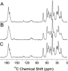
13C Ramp-CP NMR spectra of cartilage obtained under MAS after enzymatic removal of proteoglycan in (A) hydrated, (B) dehydrated, and (C) rehydrated states.

Similar articles

Cited by

References

    1. Cohen NP, Foster RJ, Mow VC. J. Orthop. Sports Phys. Ther. 1998;28:203–15. - PubMed
    1. Kirk TB, Wilson AS, Stachowiak GW. J. Orthop. Rheumatol. 1993;6:21–28.
    1. Kumar P, Oka M, Toguchida J, Kobayashi M, Uchida E, Nakamura T, Tanaka K. J. Anat. 2001;199:241–50. - PMC - PubMed
    1. Naji L, Kaufmann J, Huster D, Schiller J, Arnold K. Carbohydr. Res. 2000;327:439–46. - PubMed
    1. Huster D, Schiller J, Arnold K. Magn. Reson. Med. 2002;48:624–32. - PubMed

Publication types