. 2011 Jul 21;135(3):035103.
doi: 10.1063/1.3610427.

Does amino acid sequence determine the properties of Aβ dimer?

Affiliations

Does amino acid sequence determine the properties of Aβ dimer?

Christopher Lockhart et al. J Chem Phys. .

Abstract

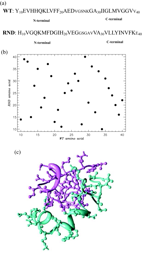
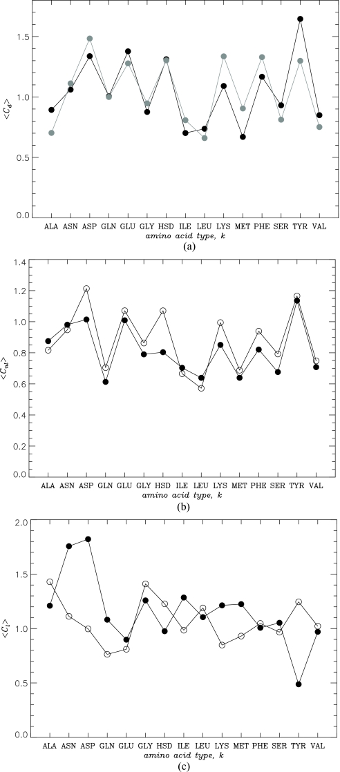
The effect of random reshuffling of amino acids on the properties of dimers formed by Aβ peptides is studied using replica exchange molecular dynamics and united atom implicit solvent model. We show that thermodynamics of dimer assembly and the dimer globule-like state are not affected by sequence permutation. Furthermore, sequence reshuffling does not change the distributions of non-local interactions and, to a large extent, amino acids in the dimer volume. To rationalize these results, we demonstrate that Gaussian statistics applies surprisingly well to the end-to-end distances of the peptides in the dimer implying that non-bonded interactions between distant along the chain amino acids are effectively screened. This observation suggests that peptides in the dimer behave as ideal chains in polymer melt, in which amino acids lose their "identity" and therefore the memory of sequence position. As a result large-scale properties of the dimer become universal or sequence independent. Comparison of our simulations with the prior theoretical studies and their implications for experiments are discussed.


Figures

Figure 1
Figure 1
(a) The sequence of wild-type (WT) Aβ10 − 40 peptide and its random (RND) mutant, in which all amino acids are randomly reshuffled. Note that although the sequence of amino acids in the WT and RND is different, their amino acid compositions are identical. Keeping the composition unchanged allows us to probe the role of sequence in dimer assembly. Amino acids from the N- and C-terminals are shown by large font. (b) Mutation map shows new positions of the WT amino acids in RND sequence. Reshuffling places amino acids at new randomly selected sequence positions. (c) Typical structure of the heterodimer composed of the WT (in purple) and RND (in aqua) peptides sampled at 360 K. The structure is visualized using Chimera (see Ref. 50)
Figure 2
Figure 2
(a) The assembly of heterodimer (HT, thick black line) and homodimer (HM, thick grey line) is compared using the number of interpeptide side chain contacts 〈Cd〉 as a function of temperature. Intrapeptide interactions are probed by the number of intrapeptide hydrogen bonds, 〈Nihb〉, as a function of temperature, in the WT (dashed black line) and RND (dashed grey line) peptides of the heterodimer. (b) Free energy F(Ch) as a function of the number of interpeptide hydrophobic contacts, Ch, computed for the heterodimer (data in black) and homodimer (data in grey) at 360 K. The free energy of dimer formation is ΔFAD = FAFD, where FA and FD = 0 are the free energies of the associated (A) and dissociated (D, Ch = 0) states. FA is obtained by integrating over the states, for which F(Ch)≤Fmin+1.0RT, where Fmin is the minimum in F(Ch). Inset: Temperature dependence of the heterodimer free energy F(T) (in black). (Note that F(T) includes the contributions from interpeptide and intrapeptide interactions and conformational entropy, but omits the ideal gas term associated with translational entropy.) Quadratic fitting function F(T) ≃ −α(TTo)2 with α=−0.003 kcal∕(mol K2), To=369 K is represented by black continuous line. For comparison, F(T) for the homodimer (see Ref. 29) is shown by grey circles. Maximum values of F(T) are set to zero. The figure demonstrates that random reshuffling of amino acids has little impact on the thermodynamics of dimer formation.
Figure 3
Figure 3
(a) Normalized radial distribution function for the number density G(r) in the heterodimer (in black) and homodimer (in grey). The distance r is measured from the dimer center of mass. G(0) represents the number density in the center of the heterodimer or homodimer. Inset: the number density pair correlation function g(r) (circles) computed for the heterodimer. Correlation radius λ is estimated by applying the exponential fit g(r)=g0 exp(−(rr0)∕λ) at r > 5 Å (continuous line). The fitting parameters are go = 0.03 Å−3, r0 = 5.3 Å, and λ = 7.3 Å. The pair correlation function g(r) computed excluding adjacent amino acids is shown by squares. For comparison, the parameters of the exponential fit to the homodimer g(r) are very similar (go = 0.03 Å−3, r0 = 5.2 Å, and λ = 7.2 Å). (b) Probability distribution P(r1N) of the distances r1N between the N- and C-terminals of the heterodimer WT peptide. Gaussian function (Eq. 1) shown by the black curve provides an excellent fit to P(r1N). Inset: the peptide segment end-to-end distance rij2 as a function of the number of amino acids in the segment |ij|. Filled and open circles are for the WT and RND peptides. Linear fits rij2=l2|ij| are shown by thick lines (l = 1.45 Å and 2.19 Å for the WT and RND peptides, respectively). Panels (a, b) suggest that the heterodimer has globule-like properties. (c) Probability Pc(k) for amino acid type k to occur in the dimer core: WT peptide (filled circles), RND peptide (open circles). Similarities in Pc(k) for the two peptides suggest that spatial distribution of amino acids in the dimer is not affected by sequence reshuffling. All plots are obtained at 360 K.
Figure 4
Figure 4
(a) The number of interpeptide side chain contacts 〈Cd(k)〉 formed by amino acids of the type k: heterodimer (HT, in black), homodimer (HM, in grey). (b,c) The numbers of non-local 〈Cnl(k)〉 (b) and local 〈Cl(k)〉 (c) intrapeptide contacts formed by amino acids of the type k in the WT (filled circles) and RND (open circles) peptides. The figure demonstrates that in contrast to local interactions amino acid sequence does not affect interpeptide and non-local intrapeptide interactions. All plots are obtained at 360 K.
Figure 5
Figure 5
(a) The fractions of β-strand 〈S(i)〉 and helix 〈H(i)〉 structure formed by amino acids i in the WT (filled circles) and RND (open circles) peptides. (b) The fractions of β-strand 〈S(k)〉 and helix 〈H(k)〉 structure formed by amino acid types k in the WT (filled circles) and RND (open circles) peptides. The figure shows that amino acid sequence not only determines the distribution of secondary structure but also the propensities of amino acid types k to adopt helix conformations. All plots are obtained at 360 K.

Similar articles

Cited by

References

    1. Dobson C. M., Nature (London) 426, 884 (2003).10.1038/nature02261 - DOI - PubMed
    1. Chiti F., Stefani M., Taddei N., Ramponi G., and Dobson C. M., Nature (London) 424, 805 (2003).10.1038/nature01891 - DOI - PubMed
    1. Song Y., Challa S. R., Medforth C. J., Qiu Y., Watt R. K., Pea D., Miller J. E., van Swolab F., and Shelnutt J. A., Chem. Commun., 1044 (2004).10.1039/b402126f - DOI - PubMed
    1. Reches M. and Gazit E., Phys. Biol. 3, S10 (2006).10.1088/1478-3975/3/1/S02 - DOI - PubMed
    1. Selkoe D. J., Nature (London) 426, 900 (2003).10.1038/nature02264 - DOI - PubMed

Publication types

Substances