. 2012 Jan;18(1-2):127-36.
doi: 10.1089/ten.TEA.2010.0290. Epub 2011 Sep 16.

Fluid shear stress alters the hemostatic properties of endothelial outgrowth cells

Affiliations

Fluid shear stress alters the hemostatic properties of endothelial outgrowth cells

Ann E Ensley et al. Tissue Eng Part A. 2012 Jan.

Abstract

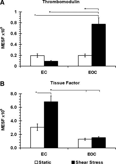
Surface endothelialization is an attractive means to improve the performance of small diameter vascular grafts. While endothelial outgrowth cells (EOCs) are considered a promising source of autologous endothelium, the ability of EOCs to modulate coagulation-related blood activities is not well understood. The goal of this study was to assess the role of arterial flow conditions on the thrombogenic phenotype of EOCs. EOCs derived from baboon peripheral blood, as well as mature arterial endothelial cells from baboons, were seeded onto adsorbed collagen, then exposed to physiologic levels of fluid shear stress. For important hemostatic pathways, cellular responses to shear stress were characterized at the gene and protein level and confirmed with a functional assay for activated protein C (APC) activity. For EOCs, fluid shear stress upregulated gene and protein expression of anticoagulant and platelet inhibitory factors, including thrombomodulin, tissue factor pathway inhibitor, and nitric oxide synthase 3 (eNOS). Fluid shear stress significantly altered the functional activity of EOCs by increasing APC levels. This study demonstrates that fluid shear stress is an important determinant of EOC hemostatic properties. Accordingly, manipulation of EOC phenotype by mechanical forces may be important for the development of thrombo-resistant surfaces on engineered vascular implants.


Figures

FIG. 1.
FIG. 1.
(A) The mononuclear fraction of blood plated onto fibronectin-coated tissue culture polystyrene following 3 days in culture is shown using phase contrast microscopy. (B, C) Endothelial progenitor outgrowth colonies formed after 9–23 days in culture and grew to confluence showing a cobblestone-like morphology. Scale bar=50 μm. (D) Representative flow cytometry histograms (EOC, passage 3) show analysis of cultured EOC protein expression. Black outlines the unstained negative control. Solid histograms are the specific antibody labeled cell samples. (E) Quantification of flow cytometry analysis. Cells with fluorescence values >90% of the negative control were considered positive. Data are expressed as mean±standard deviation, n=3. EOC, endothelial outgrowth cell; vWF, von Willebrand factor; VEGFR2, vascular endothelial growth factor receptor 2; eNOS, nitric oxide synthase 3. Color images available online at www.liebertonline.com/tea
FIG. 2.
FIG. 2.
(A) Rhodamine phalloidin staining of F-actin in cells exposed to steady laminar shear stress (15 dynes/cm2, 24 h) compared with static controls. Hoechst 33258 was used as a nuclear counterstain. Scale bar=50 μm. (B) Shape index and (C) angle of orientation were quantified (mean±SEM, n≥40). * indicates p<0.05 (ANOVA, Tukey post hoc). SEM, standard error of the mean; ANOVA, analysis of variance; EC, endothelial cell. Color images available online at www.liebertonline.com/tea
FIG. 3.
FIG. 3.
Thrombomodulin, nitric oxide synthase 3 (eNOS), tissue factor pathway inhibitor (TFPI), tissue factor (F3), and vWF gene expression in ECs and EOCs was determined by quantitative real-time reverse transcriptase–polymerase chain reaction. mRNA expression was quantified in cells exposed to steady laminar shear stress (15 dynes/cm2, 24 h) and compared with static controls (mean±SEM, n=9). * indicates p<0.05 (ANOVA, Tukey post hoc).
FIG. 4.
FIG. 4.
(A) Thrombomodulin and (B) tissue factor cell surface protein expression in EC and EOC determined by flow cytometry. Cells exposed to steady laminar shear stress (15 dynes/cm2, 24 h) were compared with static controls (mean±SEM, n≥9). * indicates p<0.05 (ANOVA, Tukey post hoc).
FIG. 5.
FIG. 5.
Generation of activated protein C (APC) by EOCs under static and steady laminar shear stress (15 dynes/cm2, 24 h) conditions. Significantly more APC was generated per cell under shear stress conditions compared with static controls (mean±SEM, n=8). * indicates p<0.05 (paired t-test).

Similar articles

Cited by

References

    1. Meinhart J.G. Deutsch M. Fischlein T. Howanietz N. Froschl A. Zilla P. Clinical autologous in vitro endothelialization of 153 infrainguinal eptfe grafts. Ann Thorac Surg. 2001;71:S327. - PubMed
    1. Deutsch M. Meinhart J. Zilla P. Howanietz N. Gorlitzer M. Froeschl A. Stuempflen A. Bezuidenhout D. Grabenwoeger M. Long-term experience in autologous in vitro endothelialization of infrainguinal eptfe grafts. J Vasc Surg. 49:352. discussion 362, 2009. - PubMed
    1. Hinds M.T. Ma M. Tran N. Ensley A.E. Kladakis S.M. Vartanian K.B. Markway B.D. Nerem R.M. Hanson S.R. Potential of baboon endothelial progenitor cells for tissue engineered vascular grafts. J Biomed Mater Res A. 2008;86:804. - PubMed
    1. Lin Y. Weisdorf D.J. Solovey A. Hebbel R.P. Origins of circulating endothelial cells and endothelial outgrowth from blood. J Clin Invest. 2000;105:71. - PMC - PubMed
    1. Melero-Martin J.M. Khan Z.A. Picard A. Wu X. Paruchuri S. Bischoff J. In vivo vasculogenic potential of human blood-derived endothelial progenitor cells. Blood. 2007;109:4761. - PubMed

Publication types

MeSH terms

LinkOut - more resources