

Effect of α-linolenic acid on endoplasmic reticulum stress-mediated apoptosis of palmitic acid lipotoxicity in primary rat hepatocytes
- PMID: 21787405
- PMCID: PMC3152932
- DOI: 10.1186/1476-511X-10-122
Effect of α-linolenic acid on endoplasmic reticulum stress-mediated apoptosis of palmitic acid lipotoxicity in primary rat hepatocytes
Abstract
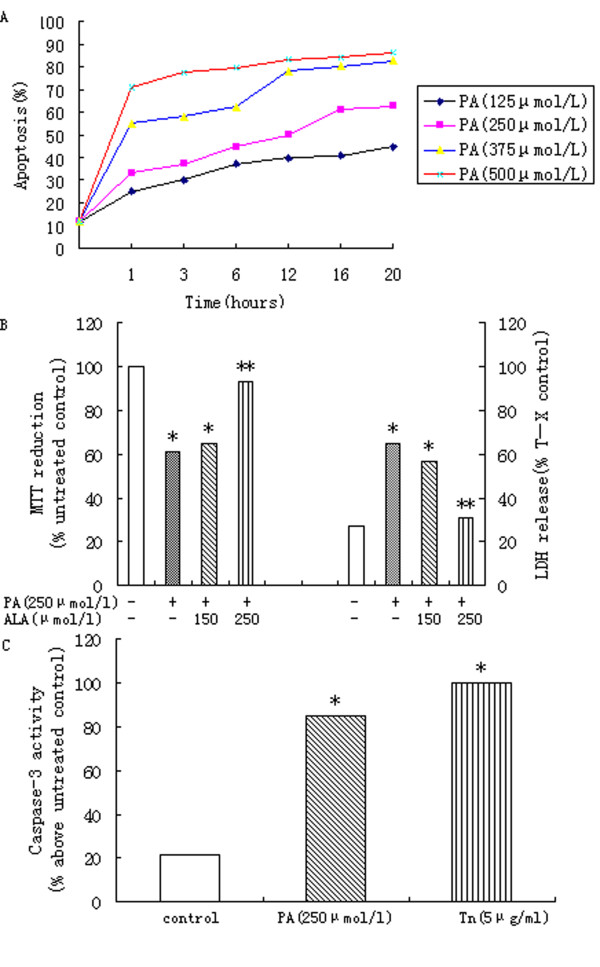
Background: Hepatic inflammation and degeneration induced by lipid depositions may be the major cause of nonalcoholic fatty liver disease (NAFLD). In this study, we investigated the effects of saturated and unsaturated fatty acids (FA) on apoptosis in primary rat hepatocytes.
Methods: The primary rat hepatocytes were treated with palmitic acid and/or α-linolenic acid in vitro. The expression of proteins associated with endoplasmic reticulum (ER) stress, apoptosis, caspase-3 levels were detected after the treatment.
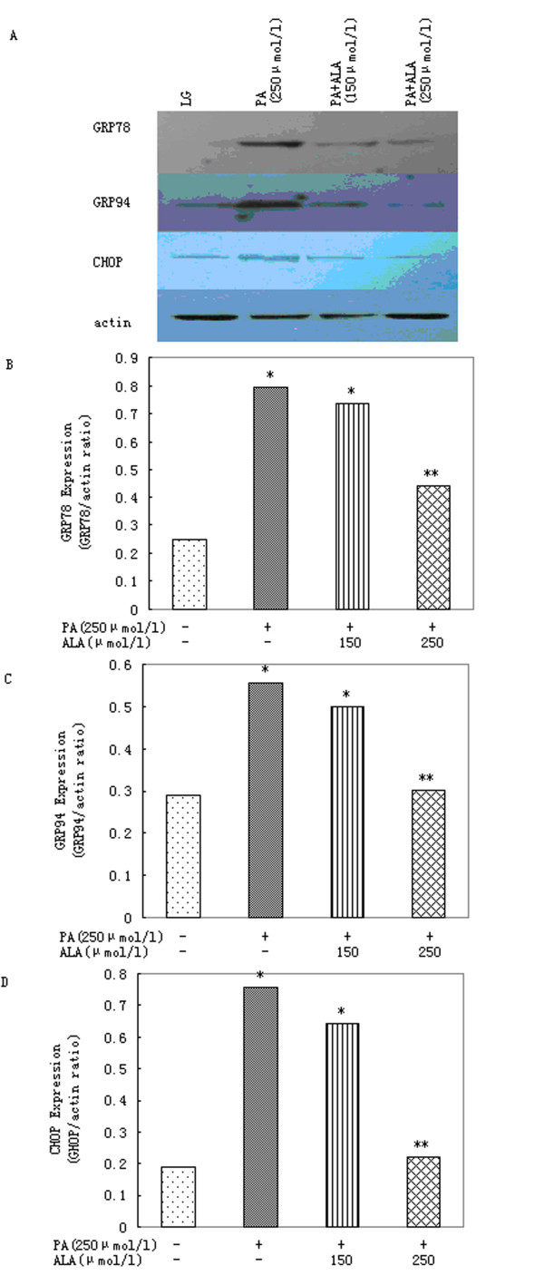
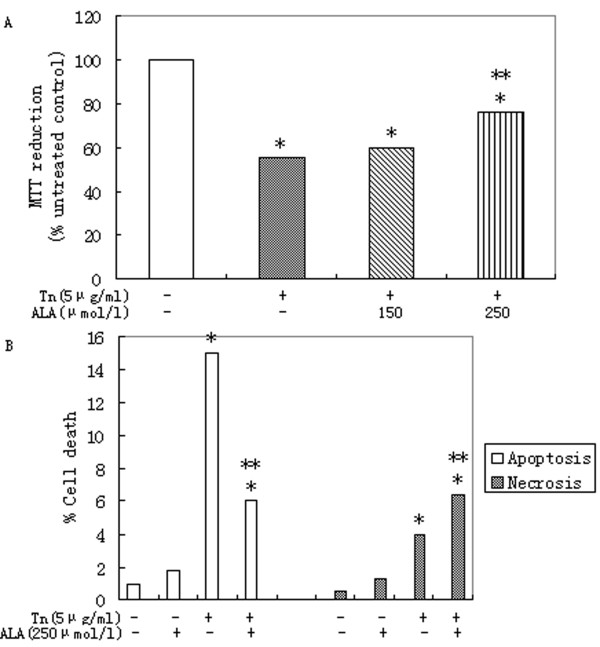
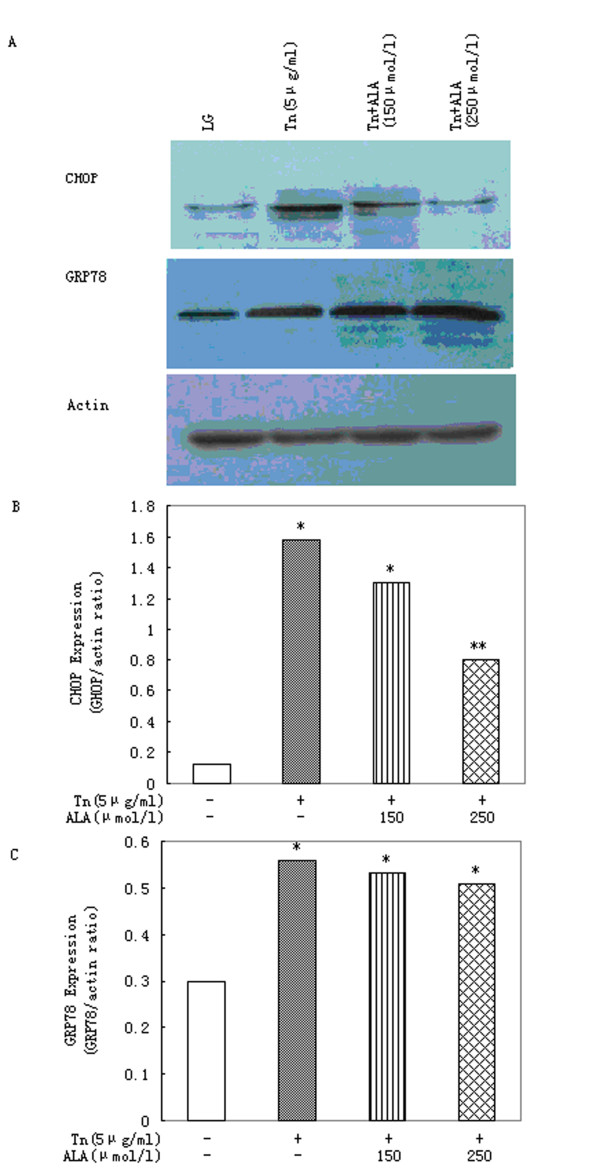
Results: The treatment with palmitic acid produced a significant increase in cell death. The unfolded protein response (UPR)-associated genes CHOP, GRP78, and GRP94 were induced to higher expression levels by palmitic acid. Co-treatment with α-linolenic acid reversed the apoptotic effect and levels of all three indicators of ER stress exerted by palmitic acid. Tunicamycin, which induces ER stress produced similar effects to those obtained using palmitic acid; its effects were also reversed by α-linolenic acid.
Conclusions: α-Linolenic acid may provide a useful strategy to avoid the lipotoxicity of dietary palmitic acid and nutrient overload accompanied with obesity and NAFLD.
Figures
Similar articles
-
α-Linolenic acid prevents endoplasmic reticulum stress-mediated apoptosis of stearic acid lipotoxicity on primary rat hepatocytes.Lipids Health Dis. 2011 May 18;10:81. doi: 10.1186/1476-511X-10-81. Lipids Health Dis. 2011. PMID: 21592363 Free PMC article.
-
alpha-Linolenic acid protects renal cells against palmitic acid lipotoxicity via inhibition of endoplasmic reticulum stress.Eur J Pharmacol. 2009 Nov 25;623(1-3):107-12. doi: 10.1016/j.ejphar.2009.09.015. Epub 2009 Sep 15. Eur J Pharmacol. 2009. PMID: 19765573
-
Palmitic and linoleic acids induce ER stress and apoptosis in hepatoma cells.Lipids Health Dis. 2012 Jan 5;11:1. doi: 10.1186/1476-511X-11-1. Lipids Health Dis. 2012. PMID: 22221411 Free PMC article.
-
Protection Strategies Against Palmitic Acid-Induced Lipotoxicity in Metabolic Syndrome and Related Diseases.Int J Mol Sci. 2025 Jan 18;26(2):788. doi: 10.3390/ijms26020788. Int J Mol Sci. 2025. PMID: 39859502 Free PMC article. Review.
-
Fatty acids and the endoplasmic reticulum in nonalcoholic fatty liver disease.Biofactors. 2011 Jan-Feb;37(1):8-16. doi: 10.1002/biof.135. Epub 2010 Dec 2. Biofactors. 2011. PMID: 21328622 Free PMC article. Review.
Cited by
-
Impact of Long-Term Supplementation with Fish Oil in Individuals with Non-Alcoholic Fatty Liver Disease: A Double Blind Randomized Placebo Controlled Clinical Trial.Nutrients. 2020 Nov 2;12(11):3372. doi: 10.3390/nu12113372. Nutrients. 2020. PMID: 33147705 Free PMC article. Clinical Trial.
-
Mechanism of Hepatocyte Apoptosis.J Cell Death. 2016 Dec 29;9:19-29. doi: 10.4137/JCD.S39824. eCollection 2016. J Cell Death. 2016. PMID: 28058033 Free PMC article. Review.
-
Molecular mechanisms of lipotoxicity and glucotoxicity in nonalcoholic fatty liver disease.Metabolism. 2016 Aug;65(8):1049-61. doi: 10.1016/j.metabol.2016.02.014. Epub 2016 Mar 3. Metabolism. 2016. PMID: 26997538 Free PMC article. Review.
-
Key fatty acid combinations define vascular smooth muscle cell proliferation and viability.Lipids. 2012 Nov;47(11):1073-84. doi: 10.1007/s11745-012-3718-6. Epub 2012 Oct 18. Lipids. 2012. PMID: 23077001
-
Omega-3 Fatty Acids and Insulin Resistance: Focus on the Regulation of Mitochondria and Endoplasmic Reticulum Stress.Nutrients. 2018 Mar 14;10(3):350. doi: 10.3390/nu10030350. Nutrients. 2018. PMID: 29538286 Free PMC article. Review.
References
-
- Solis Herruzo JA, Garcia Ruiz I, Perez Carreras M, Munoz Yague MT. Non-alcoholic fatty liver disease. From insulin resistance to mitochondrial dysfunction. Rev Esp Enferm Dig. 2006;98(11):844–874. - PubMed
MeSH terms
Substances
LinkOut - more resources
Full Text Sources
Research Materials
Miscellaneous
