. 2011 Jul 25:5:326.
doi: 10.1186/1752-1947-5-326.

Desmoid tumor of the abdominal wall: a case report

Affiliations

Desmoid tumor of the abdominal wall: a case report

Athanasios Economou et al. J Med Case Rep. .

Abstract

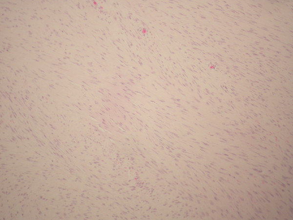
Introduction: Desmoid tumors are rare lesions without any metastatic potential but a strong tendency to invade locally and to recur. These tumors are associated with women of fertile age, especially during and after pregnancy.

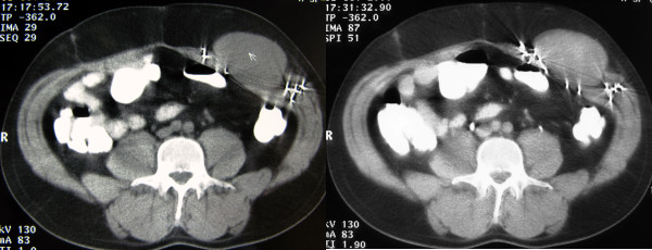
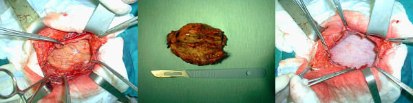
Case presentation: The case of a desmoid tumor of the anterior abdominal wall in a 40-year-old Caucasian man with no relevant family history is presented, describing its appearance on computed tomography and ultrasonography. The patient, who presented with a painless mass in the left anterolateral abdomen, had a history of previous urgent abdominal surgery after a shotgun injury two years earlier. Radical resection of the affected abdominal wall musculature was performed, and the defect was reconstructed with polypropylene mesh.

Conclusion: The diagnosis of desmoid tumor should be strongly considered even in male patients with an abdominal mass and a history of previous abdominal surgery. The goal of its treatment is complete tumor excision and avoidance of the development of complications such as hernia.


Figures

Figure 1
Figure 1
Ultrasound image showing a large mass of heterogeneous echogenicity with smooth, sharply defined margins.
Figure 2
Figure 2
Axial CT scan images of the abdomen. Computed tomography examination before (A) the intravenous administration of contrast medium revealing a well-circumscribed mass originating from the left rectus abdominis muscle and of attenuation equal to that of the muscle. The mass demonstrates a mild enhancement even in the delayed images after the intravenous administration of contrast medium (B). The presence of multiple foreign bodies caused by the shotgun injury.
Figure 3
Figure 3
Intraoperative pictures of surgery for abdominal wall desmoid tumor. A) Abdominal wall with tumor. B) Macroscopic view of the tumor. C) Abdominal wall after polypropylene mesh repair.
Figure 4
Figure 4
Microscopic view of the excised rectus desmoid tumor showing fascicles of fibroblastic spindle cells with abundant intercellular collagen. (Hematoxylin and eosin stain; original magnification × 200.)

References

    1. Teo HEL, Peh WCG, Shek TWH. Case 84: desmoid tumor of the abdominal wall. Radiology. 2005;236:81–84. doi: 10.1148/radiol.2361031038. - DOI - PubMed
    1. Arshad AR, Normala B. Surgical management of large desmoid tumour of the anterior abdominal wall. Asian J Surg. 2008;31:90–95. doi: 10.1016/S1015-9584(08)60065-2. - DOI - PubMed
    1. Stojadinovic A, Hoos A, Karpoff HM, Leung DHY, Antonescu CR, Brennan MF. et al.Soft tissue tumors of the abdominal wall. Analysis of disease patterns and treatment. Arch Surg. 2001;136:70–79. doi: 10.1001/archsurg.136.1.70. - DOI - PubMed
    1. Kumar V, Khanna S, Khanna AK, Khanna R. Desmoid tumors: experience of 32 cases and review of the literature. Indian J Cancer. 2009;46:34–39. doi: 10.4103/0019-509X.48593. - DOI - PubMed
    1. Overhaus M, Decker P, Fischer HP, Textor HJ, Hirner A. Desmoid tumors of the abdominal wall: a case report. World J Surg Oncol. 2003;1:11. doi: 10.1186/1477-7819-1-11. - DOI - PMC - PubMed

LinkOut - more resources