. 2011 Jul 25:11:166.
doi: 10.1186/1471-2180-11-166.

Campylobacter jejuni dsb gene expression is regulated by iron in a Fur-dependent manner and by a translational coupling mechanism

Affiliations

Campylobacter jejuni dsb gene expression is regulated by iron in a Fur-dependent manner and by a translational coupling mechanism

Anna D Grabowska et al. BMC Microbiol. .

Erratum in

  • BMC Microbiol. 2012;12:58

Abstract

Background: Many bacterial extracytoplasmic proteins are stabilized by intramolecular disulfide bridges that are formed post-translationally between their cysteine residues. This protein modification plays an important role in bacterial pathogenesis, and is facilitated by the Dsb (disulfide bond) family of the redox proteins. These proteins function in two parallel pathways in the periplasmic space: an oxidation pathway and an isomerization pathway. The Dsb oxidative pathway in Campylobacter jejuni is more complex than the one in the laboratory E. coli K-12 strain.

Results: In the C. jejuni 81-176 genome, the dsb genes of the oxidative pathway are arranged in three transcriptional units: dsbA2-dsbB-astA, dsbA1 and dba-dsbI. Their transcription responds to an environmental stimulus - iron availability - and is regulated in a Fur-dependent manner. Fur involvement in dsb gene regulation was proven by a reporter gene study in a C. jejuni wild type strain and its isogenic fur mutant. An electrophoretic mobility shift assay (EMSA) confirmed that analyzed genes are members of the Fur regulon but each of them is regulated by a disparate mechanism, and both the iron-free and the iron-complexed Fur are able to bind in vitro to the C. jejuni promoter regions. This study led to identification of a new iron- and Fur-regulated promoter that drives dsbA1 gene expression in an indirect way. Moreover, the present work documents that synthesis of DsbI oxidoreductase is controlled by the mechanism of translational coupling. The importance of a secondary dba-dsbI mRNA structure for dsbI mRNA translation was verified by estimating individual dsbI gene expression from its own promoter.

Conclusions: The present work shows that iron concentration is a significant factor in dsb gene transcription. These results support the concept that iron concentration - also through its influence on dsb gene expression - might control the abundance of extracytoplasmic proteins during different stages of infection. Our work further shows that synthesis of the DsbI membrane oxidoreductase is controlled by a translational coupling mechanism. The dba expression is not only essential for the translation of the downstream dsbI gene, but also Dba protein that is produced might regulate the activity and/or stability of DsbI.
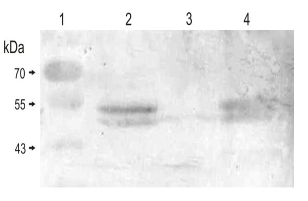

Figures

Figure 1
Figure 1
Organization of dsb genes in the C. jejuni 81-76 chromosome and constructs prepared for dsb transcription studies; the dsbA2-dsbB-astA-dsbA1 gene set (A), the dba-dsbI gene set (B). Hazy grey boxes stand for C. jejuni genes (C. jejuni NCTC 11168 and 81-176 gene numbering is given above the boxes, below them the studied gene names are given). Black boxes stand for the C. jejuni 81-176 DNA fragments cloned in the transcriptional fusions with the promoterless lacZ gene, displayed by the light grey boxes. The longest transcriptional fusion could not be obtained. Sign β-gal +/- at the right side of the plasmid name stands for presence/absence of β-galactosidase activity conferred by the appropriate construct for the transformant cells.
Figure 2
Figure 2
Transcription levels of C. jejuni 81-76 dsb genes (measured by β-galactosidase activity assays) in the wild type strain (A and B) and fur::cat mutant (C) under different environmental conditions. Each experiment was repeated three times, and each time three independent samples were taken for each strain (giving 9 independent measurements for each strain). Statistical significance was calculated using t-Student test for comparison of independent groups (GraphPad Prism). The wild type strain C. jejuni 480 carrying an empty vector pMW10 was used as a control. Statistical p values: For wild type C. jejuni 480 strain: Pdba-dsbI temp. 37°C vs 42°C: p = 0,0001(*). PdsbA2-dsbB-astA temp. 37°C vs 42°C: p = 0,2020. PdsbA1 temp. 37°C vs 42°C: p = 0,1031. Pdba-dsbI MH+Fe vs MH: p = 0,0576. Pdba-dsbI MH-Fe vs MH: p < 0,0001(*). PdsbA1-dsbB-astA MH+Fe vs MH: p = 0,0007(*). PdsbA1-dsbB-astA MH-Fe vs MH: p < 0,0001(*). PdsbA1 MH+Fe vs MH: p = 0,2569. PdsbA1 MH-Fe vs MH: p < 0,0001(*). For mutant C. jejuni 480 fur::cat strain: Pdba-dsbI MH+Fe vs MH: p = 0,3683. Pdba-dsbI MH-Fe vs MH: p = 0,6796. PdsbA1-dsbB-astA MH+Fe vs MH: p = 0,3164. PdsbA1-dsbB-astA MH-Fe vs MH: p = 0,0577. PdsbA1 MH+Fe vs MH: p = 0,5228. PdsbA1 MH-Fe vs MH: p = 0,2388. P values of P < 0.05 were considered to be statistically significant; they are marked with (*).
Figure 3
Figure 3
Electrophoretic mobility shift assays of chuA, dba-dsbI, dsbA2 and dsbA1 promoter regions bound by CjFur-His6. 28 fmol of Dig-labelled PCR amplified DNA fragments: dsbA2 (333 bp - panel A and B), dsbA1 (299 bp- panel C and D), dba-dsbI (174 bp - panel E and F) and chuA (216 bp- panel G and H) were incubated with 0, 333, 1000 or 3333 nM of purified Fur protein. The concentration of CjFur-His6 used in the reactions is indicated above the lanes. Binding buffer used in four EMSA studies (panels B, D, F, H) does not contain Mn2+. Panel I presents competition gel mobility shift assay which was performed by incubation of 3333 nmol Fur-His protein with 28 fmol of the labelled promoter region upstream of dsbA2-dsbB-astA operon (dsbA2) and various concentrations of the unlabelled promoter region upstream of dsbA2-dsbB-astA operon (dsbA2*)
Figure 4
Figure 4
Translational coupling of C. jejuni dba-dsbI. Western blot (anti-rDsbI) analysis of C. jejuni protein extracts separated by 12% SDS-PAGE. Relative positions of molecular weight markers (lane 1) are listed on the left (in kilodaltons). Lanes 2-6 contain 15 μg of total proteins from: C. jejuni 81-176 wt (2), C. jejuni 81-176 AG6 (dba-dsbI::cat) (3), AG6/pUWM811 (4), AG6/pUWM812 (5) and AG6/pUWM769 (6)
Figure 5
Figure 5
Analysis of C. jejuni dsbI transcription from a dba-dsbI operon containing wild type or point mutated dba. RT-PCR analysis of dsbI (and aphA-3) transcription in C. jejuni wild type and mutant cells. Equal amounts of mRNAs isolated from C. jejuni cells were reverse-transcribed using primer KM-R1 or Cj-RT and resulting cDNA was PCR-amplified with primer pairs KM-L1 - KM-R1 (lanes 1-7) or CjNde - Cj17RM (lanes 8-14), respectively. Relative positions of DNA molecular length markers (lanes 1, 8) are listed on the left (in base pairs). Lanes 2-6 and 9-13 contain PCR products amplified on cDNAs for C. jejuni 81-176 wt (2, 9), AG6 (dba-dsbI::cat) (3, 10), AG6/pUWM811 (4, 11), AG6/pUWM812 (5, 12), AG6/pUWM769 (6, 13); lanes 7 and 14 contain PCR products amplified on RNA for AG6/pUWM769 (after DNase treatment). White arrows indicate products of expected size.
Figure 6
Figure 6
Expression of dsbI from own promoter in C. jejuni cells. Western blot (anti-rDsbI) analysis of C. jejuni protein extracts separated by 12% SDS-PAGE. Relative positions of molecular weight markers (lane 1) are listed on the left (in kilodaltons). Lanes 2-4 contain 15 μg of total proteins from: C. jejuni 81-176 AG6 (Δdba-dsbI)/pUWM1103 (2), AG6 (3) and C. jejuni 81-176 wt (4)

Similar articles

Cited by

References

    1. Young KT, Davis LM, Dirita VJ. Campylobacter jejuni: molecular biology and pathogenesis. Nat Rev Microbiol. 2007;5(9):665–679. doi: 10.1038/nrmicro1718. - DOI - PubMed
    1. Moore JE, Corcoran D, Dooley JS, Fanning S, Lucey B, Matsuda M, McDowell DA, Megraud F, Millar BC, O'Mahony R. et al.Campylobacter. Vet Res. 2005;36(3):351–382. doi: 10.1051/vetres:2005012. - DOI - PubMed
    1. Reid AN, Pandey R, Palyada K, Naikare H, Stintzi A. Identification of Campylobacter jejuni genes involved in the response to acidic pH and stomach transit. Appl Environ Microbiol. 2008;74(5):1583–1597. doi: 10.1128/AEM.01507-07. - DOI - PMC - PubMed
    1. Malik-Kale P, Parker CT, Konkel ME. Culture of Campylobacter jejuni with sodium deoxycholate induces virulence gene expression. J Bacteriol. 2008;190(7):2286–2297. doi: 10.1128/JB.01736-07. - DOI - PMC - PubMed
    1. Woodall CA, Jones MA, Barrow PA, Hinds J, Marsden GL, Kelly DJ, Dorrell N, Wren BW, Maskell DJ. Campylobacter jejuni gene expression in the chick cecum: evidence for adaptation to a low-oxygen environment. Infect Immun. 2005;73(8):5278–5285. doi: 10.1128/IAI.73.8.5278-5285.2005. - DOI - PMC - PubMed

Publication types

MeSH terms

LinkOut - more resources