. 2011 Jul 26:10:57.
doi: 10.1186/1475-2859-10-57.

Endotoxin-free purification for the isolation of bovine viral diarrhoea virus E2 protein from insoluble inclusion body aggregates

Affiliations

Endotoxin-free purification for the isolation of bovine viral diarrhoea virus E2 protein from insoluble inclusion body aggregates

Antonino S Cavallaro et al. Microb Cell Fact. .

Abstract

Background: Protein expression in Escherichia coli may result in the recombinant protein being expressed as insoluble inclusion bodies. In addition, proteins purified from E. coli contain endotoxins which need to be removed for in vivo applications. The structural protein, E2, from Bovine Viral Diarrhoea Virus (BVDV) is a major immunogenic determinant, and is an ideal candidate as a subunit vaccine. The E2 protein contains 17 cysteine residues creating difficulties in E. coli expression. In this report we outline a procedure for successfully producing soluble and endotoxin-free BVDV E2 protein from inclusion bodies (IB).

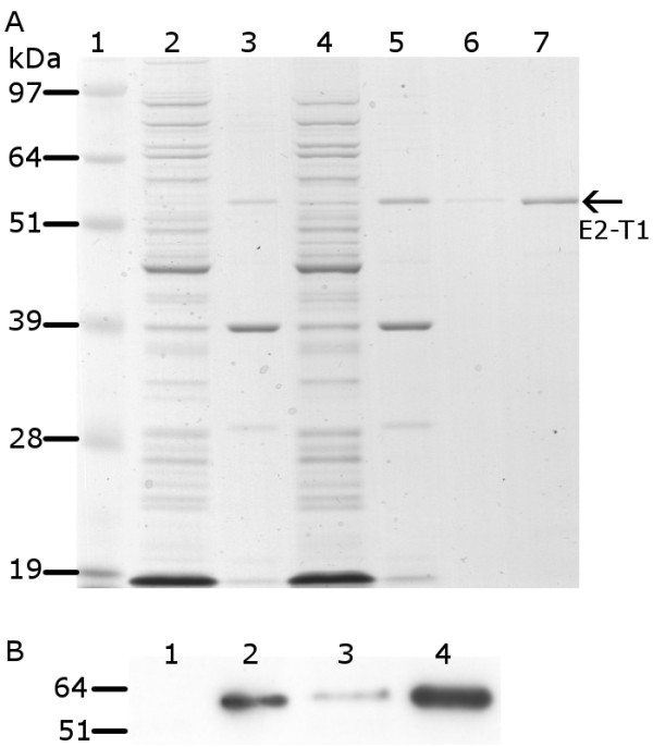
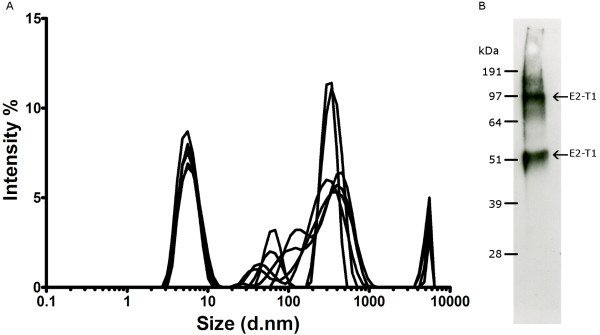
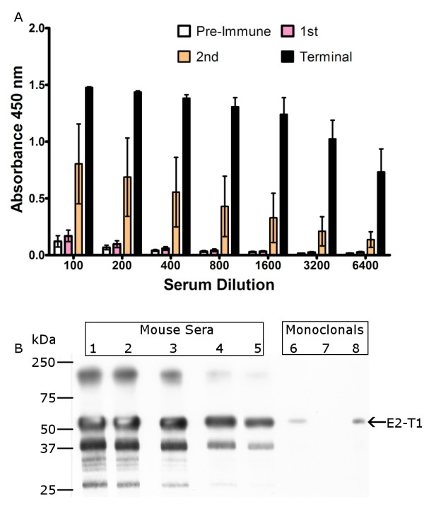
Results: The expression of a truncated form of BVDV-E2 protein (E2-T1) in E. coli resulted in predominantly aggregated insoluble IB. Solubilisation of E2-T1 with high purity and stability from IB aggregates was achieved using a strong reducing buffer containing 100 mM Dithiothreitol. Refolding by dialysis into 50 mM Tris (pH 7.0) containing 0.2% Igepal CA630 resulted in a soluble but aggregated protein solution. The novel application of a two-phase extraction of inclusion body preparations with Triton X-114 reduced endotoxin in solubilised E2-T1 to levels suitable for in vivo use without affecting protein yields. Dynamic light scattering analyses showed 37.5% of the protein was monomeric, the remaining comprised of soluble aggregates. Mice immunised with E2-T1 developed a high titre antibody response by ELISA. Western hybridisation analysis showed E2-T1 was recognised by sera from immunised mice and also by several BVDV-E2 polyclonal and monoclonal antibodies.

Conclusion: We have developed a procedure using E. coli to produce soluble E2-T1 protein from IB, and due to their insoluble nature we utilised a novel approach using Triton X-114 to efficiently remove endotoxin. The resultant protein is immunogenic and detectable by BVDV-E2 specific antibodies indicating its usefulness for diagnostic applications and as a subunit vaccine. The optimised E. coli expression system for E2-T1 combined with methodologies for solubilisation, refolding and integrated endotoxin removal presented in this study should prove useful for other vaccine applications.


Figures

Figure 1
Figure 1
Protein analysis of pET-SUMO-E2-T1 expression in E. coli BL21 (DE3). (A) Protein from soluble and insoluble fractions separated by electrophoresis on 10% Bis-Tris gel and stained with Coomassie blue. Lane 1, SeeBlue® Plus2 MW standard; lane 2, soluble protein fraction at 0 hours; lane 3, insoluble protein fraction at 0 hours; lane 4, soluble protein fraction 2 hours post IPTG induction; lane 5, insoluble protein fraction 2 hours IPTG post induction; lane 6, E2-T1 purified from IB (0.4 μL); lane 7, E2-T1 purified from IB (2 μL). (B) Western blot image of E2-T1 protein expressed in E. coli BL21 (DE3). Equivalent amounts of protein from the soluble and insoluble fractions were transferred to Hybond C and incubated with an anti-his antibody and detected by ECL. Lane 1, soluble protein fraction 0 hours; lane 2, insoluble protein fraction 0 hours; lane 3, soluble protein fraction 2 hours post IPTG induction; lane 4, insoluble protein fraction 2 hours post IPTG induction.
Figure 2
Figure 2
(A) Dynamic Light Scattering analysis of E2-T1. E2-T1 protein was dialysed into 50 mM Tris (pH 7.0) containing 0.2% Igepal CA630. The graph shows size distribution by intensity for physical size determination. Six measurements of 30 readings each were performed. (B) Western blot of purified E2-T1. E2-T1 protein was run under non-reducing conditions and probed with mouse sera raised against E2-T1.
Figure 3
Figure 3
Protein fractions following endotoxin removal from E2-T1 IB by Triton X-114 extraction were separated by electrophoresis on 10% Bis-Tris gel and stained with Coomassie blue. Lane 1, SeeBlue® Plus2 MW standards; lane 2, IB pellets resuspended in PBS before Triton X-114 treatment; lane 3, aqueous layer post Triton X-114 treatment; lane 4, recovered IB pellet post Triton X-114 treatment; lane 5, Triton TX-114 layer.
Figure 4
Figure 4
Western blot analysis of E2-T1 protein with BVDV-specific antibodies. Equivalent amounts of protein were transferred to Hybond C membrane and visualized by ECL. Lane 1, Precision Plus Protein Kaleidoscope MW standards; lane 2, Anti-His antibody; lane 3, sheep 804 pre immune sera; lane 4, sheep 804 post immune sera; lane 5, VMRD monoclonal D89; lane 6, VMRD monoclonal 157; lane 7, VMRD monoclonal 348; lane 8, Linfa Wang D4/G4 monoclonal; lane 9, VMRD Goat anti BVDV; lane 10, Anti-His antibody.
Figure 5
Figure 5
Serum analysis from E2-T1 inoculated mice. (A) ELISA analysis of mice receiving three injections of E2-T1. Mice (n = 4) were injected with 50 μg E2-T1 and 10 μg QuilA at 2 week intervals. ELISA assays were performed using pre-immune sera and sera obtained two weeks following each injection, termed 1st, 2nd and terminal samples. (B) Western blot analysis of E2-T1 with terminal sera. Mice sera was used at the following dilutions: lane 1, 1:4000; lane 2, 1:8000; lane 3, 1:16000; lane 4, 1:32000; lane 5, 1:64000; lane 6, VMRD monoclonal 348; lane 7, VMRD monoclonal D89; lane 8, VMRD monoclonal 157.

Similar articles

Cited by

References

    1. Houe H. Epidemiological features and economical importance of bovine virus diarrhoea virus (BVDV) infections. Veterinary Microbiology. 1999;64(2-3):89–107. doi: 10.1016/S0378-1135(98)00262-4. - DOI - PubMed
    1. Houe H. Economic impact of BVDV infection in dairies. Biologicals. 2003;31(2):137–143. doi: 10.1016/S1045-1056(03)00030-7. - DOI - PubMed
    1. Thiel HJ, Collett MS, Gould EA, Heinz FX, Houghton M, Meyers G, Purcell RH, Rice CM. In: Virus Taxonomy Eighth Report on the International Committee on the Taxonomy of Viruses. Fauquet CM, Mayo MA, Maniloff J, Desselberger U, Ball LA, editor. Amsterdam: Academic Press; 2005. Family Flaviviridae; pp. 981–998.
    1. Bolin SR. Immunogens of Bovine Viral Diarrhea Virus. Veterinary Microbiology. 1993;37(3-4):263–271. doi: 10.1016/0378-1135(93)90028-6. - DOI - PubMed
    1. Bolin SR, Ridpath JF. Glycoprotein E2 of bovine viral diarrhea virus expressed in insect cells provides calves limited protection from systemic infection and disease. Archives of Virology. 1996;141(8):1463–1477. doi: 10.1007/BF01718248. - DOI - PubMed

Publication types

MeSH terms

LinkOut - more resources