. 2011 Sep 9;412(1):14-21.
doi: 10.1016/j.jmb.2011.07.014. Epub 2011 Jul 22.

Crystal structure of the cytoplasmic N-terminal domain of subunit I, a homolog of subunit a, of V-ATPase

Affiliations

Crystal structure of the cytoplasmic N-terminal domain of subunit I, a homolog of subunit a, of V-ATPase

Sankaranarayanan Srinivasan et al. J Mol Biol. .

Erratum in

  • J Mol Biol. 2011 Oct 21;413(2):523

Abstract

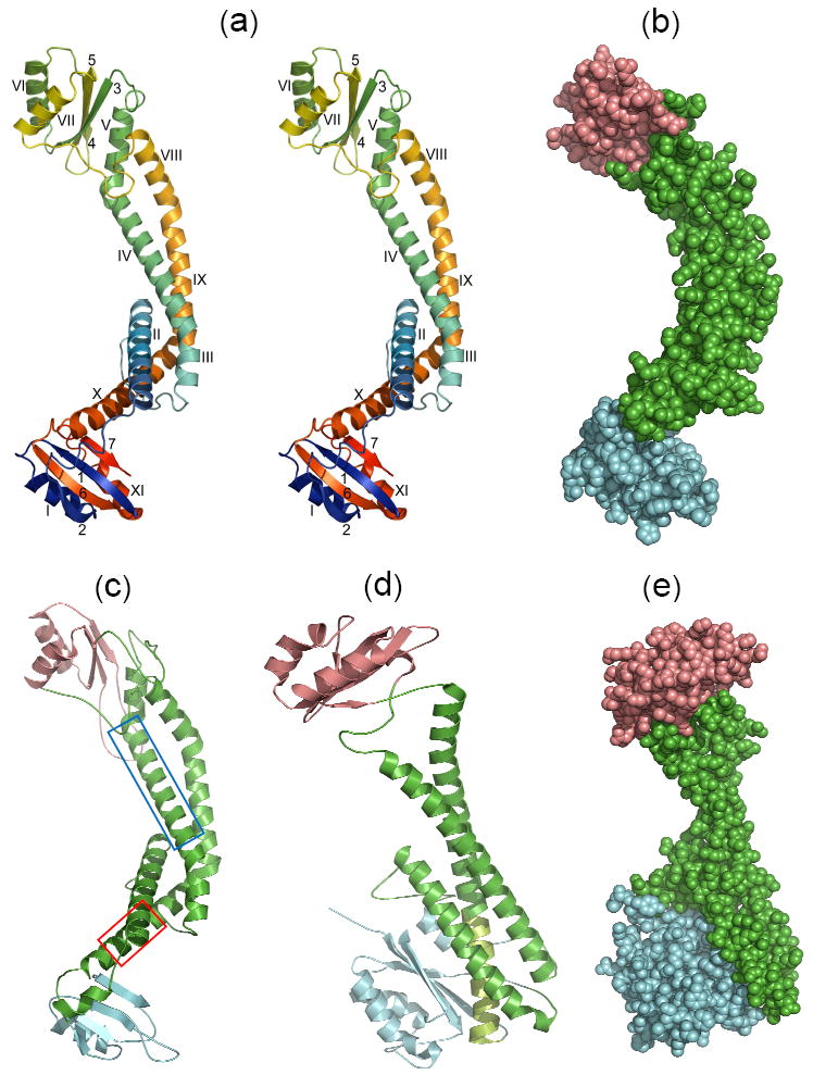
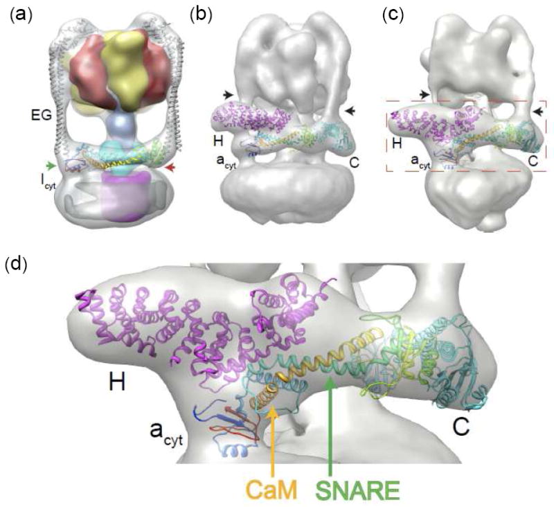
Subunit "a" is associated with the membrane-bound (V(O)) complex of eukaryotic vacuolar H(+)-ATPase acidification machinery. It has also been shown recently to be involved in diverse membrane fusion/secretory functions independent of acidification. Here, we report the crystal structure of the N-terminal cytosolic domain from the Meiothermus ruber subunit "I" homolog of subunit a. The structure is composed of a curved long central α-helix bundle capped on both ends by two lobes with similar α/β architecture. Based on the structure, a reasonable model of its eukaryotic subunit a counterpart was obtained. The crystal structure and model fit well into reconstructions from electron microscopy of prokaryotic and eukaryotic vacuolar H(+)-ATPases, respectively, clarifying their orientations and interactions and revealing features that could enable subunit a to play a role in membrane fusion/secretion.


Figures

Fig. 1
Fig. 1
X-ray and modeled structures in similar orientations. (a) Stereoview of the ribbon backbone trace of the Icyt crystal structure color ramped from the N- to the C-terminal ends, and α-helices and β-sheet strands identified with Roman and Arabic numbers, respectively. Superposition of the distal (top) and proximal (bottom) lobes showed an rmsd of 5.3 Å over 23 α-carbons. (b) Space-filling representation of Icyt structure. The long linker region of anti-parallel pairs of α-helices is colored green, and the distal and proximal lobes are distinguished by salmon and cyan colors, respectively. These distinctions are adopted in c to e. (c) Model of acyt of the neuronal zebrafish subunit a1 obtained as described in the Supplementary Methods and Table S3. The α-helices boxed in blue (αV) and red (αX) in the sequence alignment which are equivalent to Icyt’s αIV and αX, respectively, have been implicated as sites for binding of t-SNARE protein syntaxin or SNAP25 and Ca2+-calmodulin, correspondingly (described further in Supplementary Fig. S3). (d) Ribbon trace and (e) space-filling representation, of the yeast subunit C (Vma5p) crystal structure (PDB 1U7L). The match from the DALI database between Icyt and subunit C structure gave Z score of 6.8 and rmsd of 10.1 Å over 216 aligned residues and sequence identity of 7%. A large substructure is formed by extensive hydrophobic interactions of the C-terminal segment of the longest α helix (light green) of the linker with the inner face of β-sheet of the foot domain (blue).
Fig. 2
Fig. 2
Fitting of the Icyt crystal structure and acyt model to respective EM densities. The methods are described in the Supplementary Information. All the fits are unique as assessed by e3fhstat.py in EMAN2 which indicated 99th percentile for volume inclusion and real-space correlation (for additional statistics see Supplementary Methods). (a) Fit of the crystal structures of Icyt (color ramped ribbon trace) and the T. thermophilus heterodimer of subunits EG (gray trace) into the 16 Å cryo-EM density of the T. thermophilus V-ATPase. The surface density (available from ref. 15) of subunits A is colored yellow; B, red; D, blue; C, cyan; and c, magenta. The green and red arrows indicate the proximal and distal lobes of Icyt, respectively. (b) Fit of the zebrafish acyt model (Fig. 1c) color ramped trace and crystal structures of yeast subunits H20 and C14 into the 25 Å negative-stain EM reconstruction density of yeast V-ATPase. The black arrows in this figure and that in panel c indicate the vertical column of density of two of the three peripheral stalks. (c) Fit of the identical components identified in b into the 17 Å cryo-EM reconstruction density of M. sexta V-ATPase. Red dashed rectangle delineates an area of the V-ATPase fit that has been magnified in (d). In both Figs. b and c, the third peripheral stalk is in the “rear” and obstructed from view. Compared to yeast V-ATPase, in M. sexta, acyt and subunit H are rotated clockwise by ~25° and ~30°, respectively, about a horizontal axis, whereas subunit C is rotated clockwise by ~ 10° about an axis normal to the plane of the figure. This level of difference may be attributed to the differences in the imaging methods and the resulting resolutions. (d) A magnified region of the zebrafish acyt fit delineated in panel c. Potential binding sites for SNARE and Ca2+-calmodulin (CaM) are indicated by green and orange arrows, respectively.

References

    1. Forgac M. Vacuolar ATPases: rotary proton pumps in physiology and pathophysiology. Nat Rev Mol Cell Biol. 2007;8:917–929. - PubMed
    1. Muench SP, Trinick J, Harrison MA. Structural divergence of the rotary ATPases. Q Rev Biophys. 2011:1–46. - PubMed
    1. Yoshida M, Muneyuki E, Hisabori T. ATP synthase-a marvellous rotary engine of the cell. Nat Rev Mol Cell Biol. 2001;2:669–77. - PubMed
    1. Kane PM. Disassembly and reassembly of the yeast vacuolar H(+)-ATPase in vivo. J Biol Chem. 1995;270:17025–32. - PubMed
    1. Sumner JP, Dow JA, Earley FG, Klein U, Jager D, Wieczorek H. Regulation of plasma membrane V-ATPase activity by dissociation of peripheral subunits. J Biol Chem. 1995;270:5649–53. - PubMed

Publication types

MeSH terms

Associated data

LinkOut - more resources