

Wide-field functional imaging of blood flow and hemoglobin oxygen saturation in the rodent dorsal window chamber
- PMID: 21787792
- PMCID: PMC3215846
- DOI: 10.1016/j.mvr.2011.07.004
Wide-field functional imaging of blood flow and hemoglobin oxygen saturation in the rodent dorsal window chamber
Abstract
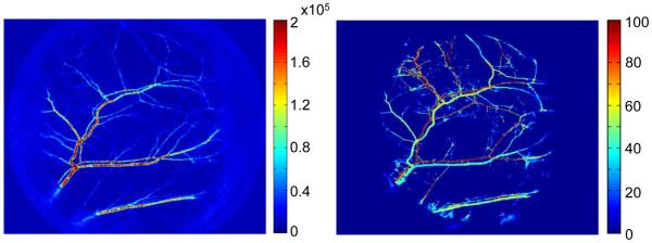
The rodent dorsal window chamber is a widely used in vivo model of the microvasculature. The model consists of a 1cm region of exposed microvasculature in the rodent dorsal skin that is immobilized by surgically implanted titanium frames, allowing the skin microvasculature to be visualized. We describe a detailed protocol for surgical implantation of the dorsal window chamber which enables researchers to perform the window chamber implantation surgery. We further describe subsequent wide-field functional imaging of the chamber to obtain hemodynamic information in the form of blood oxygenation and blood flow on a cm size region of interest. Optical imaging techniques, such as intravital microscopy, have been applied extensively to the dorsal window chamber to study microvascular-related disease and conditions. Due to the limited field of view of intravital microscopy, detailed hemodynamic information typically is acquired from small regions of interest, typically on the order of hundreds of μm. The wide-field imaging techniques described herein complement intravital microscopy, allowing researchers to obtain hemodynamic information at both microscopic and macroscopic spatial scales. Compared with intravital microscopy, wide-field functional imaging requires simple instrumentation, is inexpensive, and can give detailed metabolic information over a wide field of view.
Copyright © 2011 Elsevier Inc. All rights reserved.
Figures


References
-
- Algire GH. An adaptation of the transparent-chamber technique to the mouse. J. Natl. Cancer Inst. 1943;4:1–11.
-
- Briers J, Fercher A. Retinal blood-flow visualization by means of laser speckle photography. Invest Ophthalmol Vis Sci. 1982;22:255–9. - PubMed
-
- Briers JD, Webster S. Laser speckle contrast analysis (LASCA): a nonscanning, full-field technique for monitoring capillary blood flow. J Biomed Opt. 1996;1:174–179. - PubMed
Publication types
MeSH terms
Substances
Grants and funding
LinkOut - more resources
Full Text Sources
Other Literature Sources
Medical
