Review
. 2011 Oct;34(10):515-25.
doi: 10.1016/j.tins.2011.06.006. Epub 2011 Jul 23.

Pattern separation in the hippocampus

Affiliations
Review

Pattern separation in the hippocampus

Michael A Yassa et al. Trends Neurosci. 2011 Oct.

Abstract

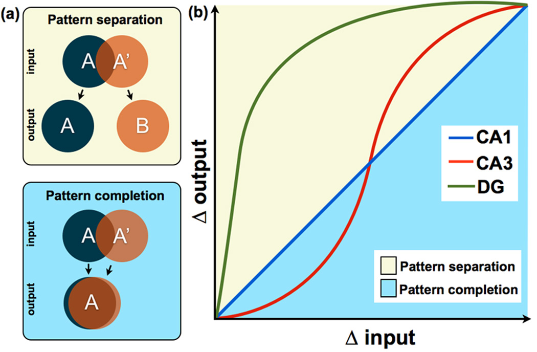
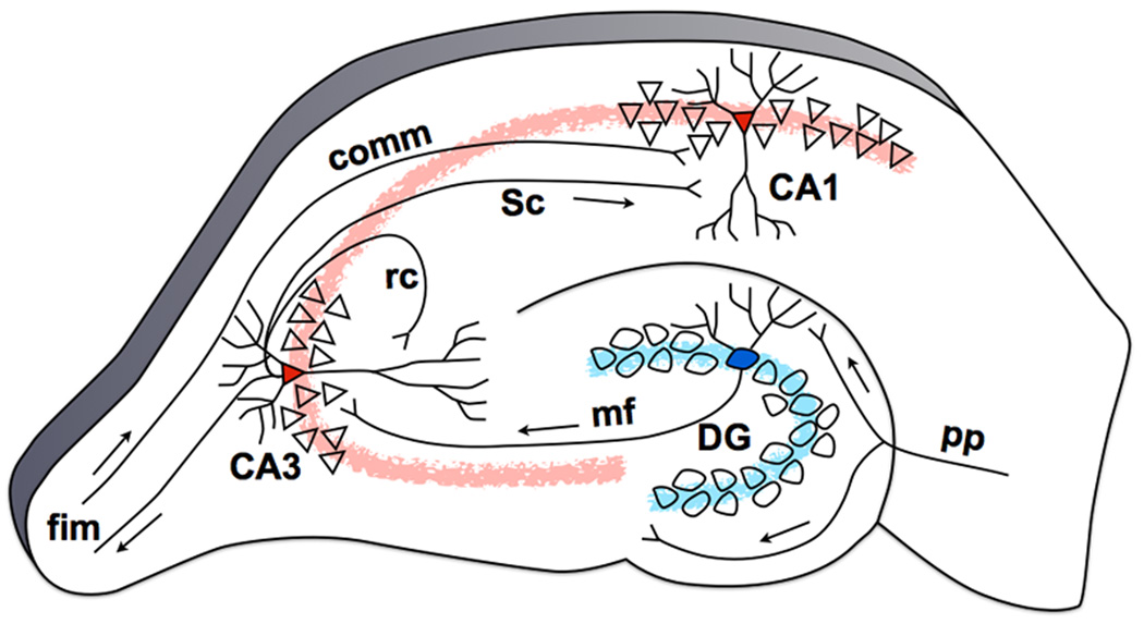
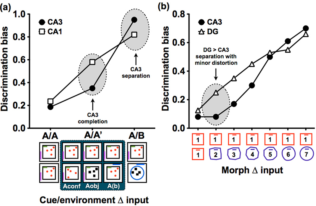
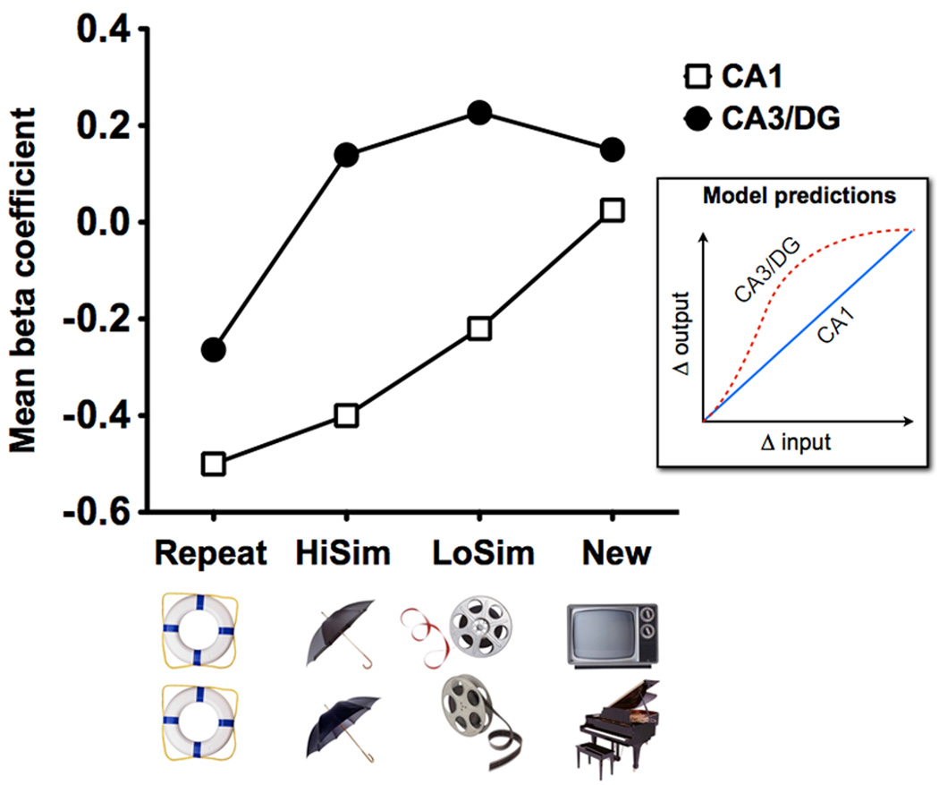
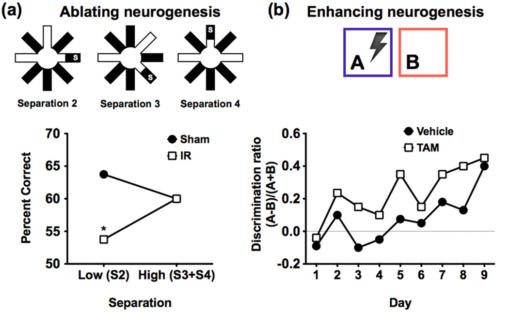
The ability to discriminate among similar experiences is a crucial feature of episodic memory. This ability has long been hypothesized to require the hippocampus, and computational models suggest that it is dependent on pattern separation. However, empirical data for the role of the hippocampus in pattern separation have not been available until recently. This review summarizes data from electrophysiological recordings, lesion studies, immediate-early gene imaging, transgenic mouse models, as well as human functional neuroimaging, that provide convergent evidence for the involvement of particular hippocampal subfields in this key process. We discuss the impact of aging and adult neurogenesis on pattern separation, and also highlight several challenges to linking across species and approaches, and suggest future directions for investigation.


Figures

Figure 1
Figure 1. Schematic representation of input/output transfer functions in hippocampal subfields
(a) A conceptual representation of pattern separation and pattern completion. Pattern separation can be thought of as making similar, overlapping representations (i.e., A and A’) more distinct, while pattern completion can be thought of as making overlapping representations even more overlapping. (b) Figure adapted from [28], showing a nonlinear transformation in CA3 but not in CA1. A curve for the DG has been added to indicate that neurons in this region respond nonlinearly to small increments of change in sensory input. The diagonal line represents the scenario where input and output are equal i.e., Δ input = Δ output), whereas the yellow portion of the plot above the diagonal describes situations in which input is made more dissimilar (i.e., separation: Δ output > Δ input). The blue portion below the diagonal describes situations in which input is made more similar (i.e., completion: Δ output < Δ input). In this scheme, pattern separation and completion are defined in terms of the extent to which a tuning function deviates from the diagonal. This schematic is based on data across many studies in animals and humans [–27,29,30,34].
Figure 2
Figure 2. The hippocampal tri-synaptic circuit based on the rat brain
Neurons in Layer II of the EC project to the DG, bypassing the subiculum, with additional collaterals projecting to the CA3 subfield (perforant path, pp). Granule cells in the DG project to the CA3 field of the hippocampus via the mossy fiber (mf) pathway. The CA3’s pyramidal cells project heavily onto themselves via recurrent collaterals (rc) and also to the CA1 through Schaffer collaterals (Sc). This trisynaptic circuit is a primarily feedforward circuit with very little feedback, except from the CA3 back to the DG via the hilar mossy cells [12] (not shown). The fimbria/fornix (fim) is one of the principal output pathways of the hippocampus that also brings in commissural (comm) input from the contralateral hippocampus.
Figure 3
Figure 3. Hippocampal subfield dynamics
(a) Figure based on data in [27], depicting circumstances that elicit separation and completion in CA3, based on a study using IEG brain imaging in rats. Discrimination bias was calculated as the inverse of the overlap scores used in the original paper. Here, the dependent measure was the degree of overlap in representations, as assessed by expression of the IEG Arc, when the test environment was the same (A/A), similar (A/A’), or distinct (A/B) from the original environment. With minor distortions of the original environment (i.e., A/A’), such as a change in object configuration (Aconf), its identity (Aobj), or a displacement of the entire maze in a different but similar room (A(b)), evidence of pattern completion was observed in CA3. However, when rats were tested in a new environment (I.e., A/B), CA3 demonstrated evidence of pattern separation. (b) Figure based on data in [29], depicting simultaneous electrophysiological recordings from CA3 and DG in rats as the test environment was incrementally morphed from the original environment (1) into a novel environment (7). Discrimination bias was once again calculated as the inverse of the overlap scores used in the original paper. Data shows higher pattern separation in the DG compared to the CA3 even with the smallest distortions in the environment.
Figure 4
Figure 4. Pattern separation in the human DG/CA3 as measured by BOLD fMRI
In an incidental encoding paradigm in which participants were asked to indicate whether each picture was of an “indoor” or an “outdoor” item, BOLD fMRI activity was used to track the similarity of objects [34] (HiSim = high similarity, LoSim = low similarity). CA3/DG activity showed evidence of pattern separation, as evidenced by a rapid, nonlinear response to even small changes in input (N.B. two regions within CA3/DG exhibited activity consistent with pattern separation and one was ambiguous; only data from the clusters showing pattern separation was averaged to produce this curve. Data from all three shown in [34]). In contrast, CA1 activity showed evidence for incremental (linear) changes consistent with the pattern predicted by the model shown in the inset. Since CA3 and DG cannot be dissociated in fMRI studies, even at high-resolution, the prediction of the model was produced by extrapolating a combined function for the DG and CA3 (see Figure 1 for more details).
Figure 5
Figure 5. The effects of manipulating neurogenesis on pattern separation
(a) In this study [80] in rats input similarity was manipulated by varying the number of intervening radial maze arms between the sample and target locations. In this delayed non-match to place paradigm, a smaller number of intervening arms (e.g. 2) should require more pattern separation than larger numbers (e.g. 3 and 4). The results show that x-irradiated mice (IR) that lacked neurogenesis were impaired at low (S2) but not high (S3 and S4) separations compared to sham test mice. These results illustrate that newborn granule cells are important for normal pattern separation. Figure adapted, with permission, from AAAS [80]. (b) In this study [81], contextual fear discrimination learning with similar environments (A and B) was used to test pattern separation abilities in a group of mice with a genetically inducible manipulation that enhances newborn neuron survival in the DG. Results show higher discrimination in the mice with enhanced neurogenesis (TAM) compared to control mice (vehicle) during the first eight days of learning. This gain-of-function manipulation illustrates that increasing the number of adult-born neurons improves pattern separation. Figure adapted, with permission, from Macmillan Publishers Ltd [81]. Together, results from (a) and (b) suggest an important role for newborn neurons in DG-mediated pattern separation.

References

    1. Marr D. Simple memory: a theory for archicortex. Philos Trans R Soc Lond B Biol Sci. 1971;262:23–81. - PubMed
    1. McClelland JL, et al. Why there are complementary learning systems in the hippocampus and neocortex: insights from the successes and failures of connectionist models of learning and memory. Psychol Rev. 1995;102:419–457. - PubMed
    1. Norman KA, O'Reilly RC. Modeling hippocampal and neocortical contributions to recognition memory: a complementary-learning-systems approach. Psychol Rev. 2003;110:611–646. - PubMed
    1. McNaughton BL, Morris RG. Hippocampal synaptic enhancement and information storage within a distributed memory system. Trends Neurosci. 1987;10:408–415.
    1. Shapiro ML, Olton DS. Hippocampal function and interference. In: Schacter DL, Tulving E, editors. Memory Systems 1994. MIT Press; 1994. pp. 141–146.

Publication types