. 2011 Sep 15;71(18):6061-72.
doi: 10.1158/0008-5472.CAN-10-4269. Epub 2011 Jul 25.

Endothelial cells create a stem cell niche in glioblastoma by providing NOTCH ligands that nurture self-renewal of cancer stem-like cells

Affiliations

Endothelial cells create a stem cell niche in glioblastoma by providing NOTCH ligands that nurture self-renewal of cancer stem-like cells

Thant S Zhu et al. Cancer Res. .

Abstract

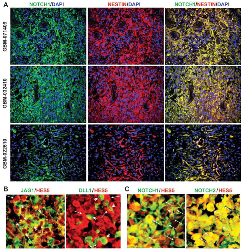
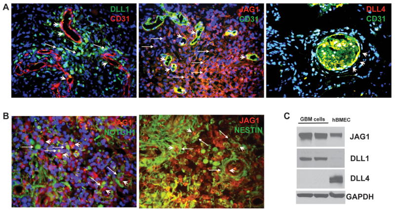
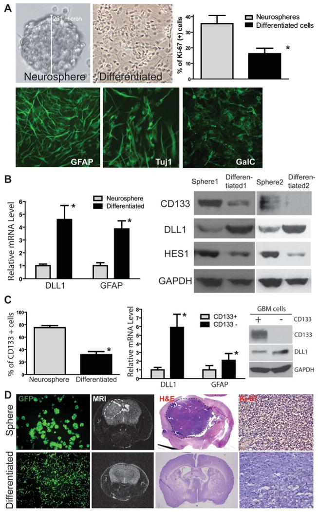
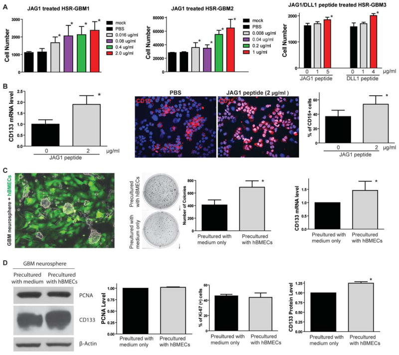
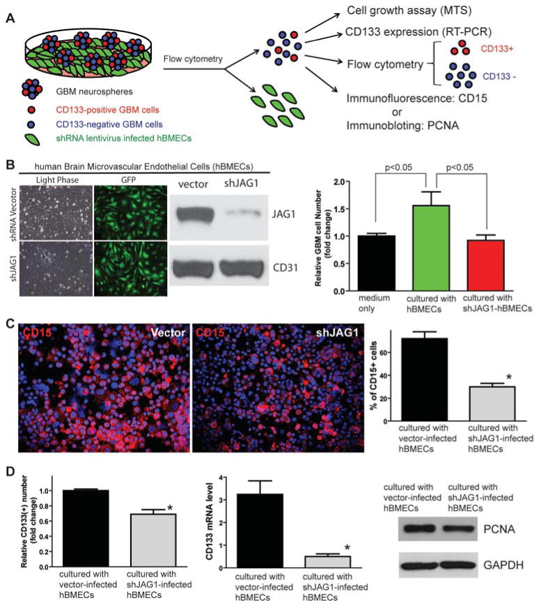
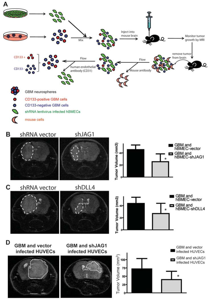
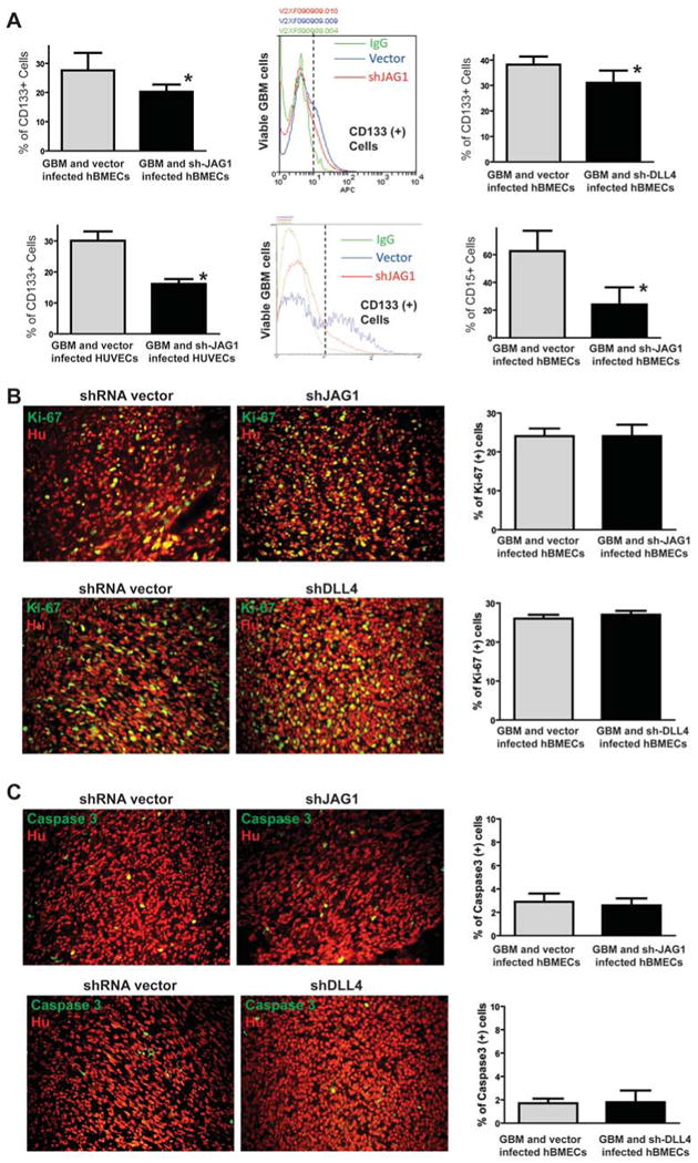
One important function of endothelial cells in glioblastoma multiforme (GBM) is to create a niche that helps promote self-renewal of cancer stem-like cells (CSLC). However, the underlying molecular mechanism for this endothelial function is not known. Since activation of NOTCH signaling has been found to be required for propagation of GBM CSLCs, we hypothesized that the GBM endothelium may provide the source of NOTCH ligands. Here, we report a corroboration of this concept with a demonstration that NOTCH ligands are expressed in endothelial cells adjacent to NESTIN and NOTCH receptor-positive cancer cells in primary GBMs. Coculturing human brain microvascular endothelial cells (hBMEC) or NOTCH ligand with GBM neurospheres promoted GBM cell growth and increased CSLC self-renewal. Notably, RNAi-mediated knockdown of NOTCH ligands in hBMECs abrogated their ability to induce CSLC self-renewal and GBM tumor growth, both in vitro and in vivo. Thus, our findings establish that NOTCH activation in GBM CSLCs is driven by juxtacrine signaling between tumor cells and their surrounding endothelial cells in the tumor microenvironment, suggesting that targeting both CSLCs and their niche may provide a novel strategy to deplete CSLCs and improve GBM treatment.


Figures

Figure 1
Figure 1. NOTCH receptor-expressing cells are co-localized with NESTIN-positive tumor cells in primary GBM and have elevated level of NOTCH activity adjacent to NOTCH-ligand expressing cells
(A) Expression of NOTCH1 and NESTIN was co-localized in the same cells in the primary GBM tumors (GBM-071409, GBM-032410, and GBM-022610). (B) JAG1-expressing cells or DLL1-expressing cells (green, arrowhead) were adjacent to HES5-expressing cells (red, arrow) in primary GBM samples. (C) NOTCH1- or NOTCH2-expressing cells were co-localized with HES5-expressing cells (arrow) in primary GBM samples.
Figure 2
Figure 2. Expression pattern of NOTCH ligands in primary GBMs
(A) NOTCH ligand DLL1 (green) was expressed in some GBM cells (arrow) and in some endothelial cells (CD31+) around the blood vessels (arrow head). JAG1 was expressed in both tumor cells (arrow) and blood vessels (arrow head). DLL4 was expressed in the blood vessels and co-localized with CD31 staining in endothelial cells (arrow head). (B) NOTCH1 receptor and CSLC marker NESTIN were expressed in tumor cells (arrow) adjacent to JAG1-expressing endothelial cells or tumor cells (arrowhead). (C) Western blots showed expression of JAG1, DLL1 and DLL4 in GBM neurosphere cells (Lane 1 and 2: same GBM cells) and hBMECs (Lane 3), consistent with immunohistochemistry results. GAPDH was used as a loading control.
Figure 3
Figure 3. Differentiated GBM cells express NOTCH ligands and have reduced ability to form intracranial xenograft in mice
(A) HSR-GBM1 neurosphere line was forced to differentiate and grow as a mono-layer, which had reduced proliferation (*: p<0.05, t-test) and induced expression of GFAP (glial), Tuj1 (neuronal) and GalC (oligodendrocyte) markers. (B) Differentiated HSR-GBM1 cells expressed higher levels of DLL1 and GFAP at the mRNA level detected by quantitative RT-PCR (left panel). In addition, differentiated cells expressed a higher level of DLL1 and lower levels of NOTCH target HES1 and stem cell marker CD133 at the protein level as detected by Western blot in two different GBM neurosphere line HSR-GBM1 and HSR-GBM2. (C) CD133-positive population was significantly reduced in differentiated tumor cells compared with GBM neurospheres (HSR-GBM1) detected by flow cytometry (left panel). In addition, CD133-negative population in HSR-GBM1 neurospheres expressed higher levels of DLL1 and GFAP at mRNA level compared with CD133-positive population (middle panel). Up-regulation of DLL1 in CD133-negative population was also confirmed at the protein level by Western blot (right panel). (D) When GBM neurospheres or differentiated cells from HSR-GBM1 labeled with GFP using lentivirus were injected into the mouse brain, neurosphere cells formed large xenografts as detected by MRI (dash circle), whereas differentiated cells had reduced ability to form xenografts. Pathology was confirmed by H&E staining and immunostaining of the proliferation marker Ki-67.
Figure 4
Figure 4. NOTCH ligand JAG1 or DLL1 promotes GBM neurosphere growth in vitro
(A) Soluble ligand JAG1 peptide treatment for 5 days induced growth of HSR-GBM1 and HSR-GBM2 in a dose-dependent fashion (left and middle panels). JAG1 or DLL1 peptide also induced growth of HSR-GBM3 (right panel). (B) 2 μg/ml JAG1 peptide treatment increased CD133 mRNA expression in HSR-GBM1 (left panel). 2 μg/ml JAG1 peptide treatment also increased the CD15-positive CSLC population in GBM neurospheres detected by immunofluorescence (right panel, n=6 random fields, *: p<0.05, t-test). (C) GBM neurospheres co-cultured with GFP-labeled hBMECs were sorted by flow cytometry and examined clonogenesis by soft agar formation assay. GBM cells formed more colonies when precultured with hBMECs (n=6 wells were counted, *: p<0.01, t-test). In addition, expression of CSLC marker CD133 was also induced at mRNA level in GBM neurospheres pre-cultured with hBMECs compared to those pre-cultured with medium only (right panel, n=6 repeats of this experiment, *: p<0.01, t-test). (D) There was no proliferation change in GBM neurospheres pre-cultured with or without hBMECs, identified by PCNA protein expression using Western blot and percentage of Ki-67 positive population by immunofluorescence. However, CD133 expression was significantly induced in GBM neurospheres pre-cultured with hBMECs (right panel). p-Actin was used as a loading control.
Figure 5
Figure 5. Knocking down JAG1 expression in hBMECs by shRNA decreases CSLC population in co-cultured GBM neurospheres in vitro
(A) Schematic showing the experimental approach used to examine if NOTCH ligands expressed in endothelial cells are essential for co-cultured GBM neurosphere growth. (B) Western blot showed that JAG1 expression was significantly knocked down by shJAG1 lentiviruses in hBMECs to be co-cultured with GBM neurospheres. CD31 was served as an internal control. HSR-GBM1 growth was increased when co-cultured with hBMECs compared to neurospheres cultured by medium only, whereas knockdown of JAG1 expression in hBMECs abrogated hBMEC-induced HSR-GBM1 growth. (C) CD15-positive CSLC population detected by immunofluorescence was also reduced in GBM neurospheres co-cultured with shJAG1-infected hBMECs compared to GBM neurospheres co-cultured with empty vector-infected hBMECs. (D) Knockdown of JAG1 expression in hBMECs decreased CD133-positive population in HSR-GBM1 neurospheres co-cultured with hBMECs compared to HSR-GBM1 neurospheres cultured with empty vector-infected hBMECs (left). mRNA expression of CD133 was also lowered in HSR-GBM1 neurosphere cells co-cultured with sh-JAG1-infected hBMECs compared to HSR-GBM1 cells co-cultured with empty vector-infected hBMECs (middle). There was no proliferation change in HSR-GBM1 neurospheres co-cultured with empty vector- or shJAG1-infected hBMECs, identified by PCNA protein expression using Western blot (right).
Figure 6
Figure 6. Knocking down JAG1 or DLL4 expression by shRNA in hBMECs inhibits growth of GBM intracranial xenografts derived from the mixture of GBM neurospheres and hBMECs
(A) Schematic showing the experimental approach used to examine if NOTCH ligand expressed in endo-thelial cells are essential for the growth of GBM intracranial xenografts derived from the mixture of GBM neurospheres and endothelial cells. (B) MRI showed that the growth of GBM intracranial xeno-grafts derived from a mixture of GBM neurospheres and shJAG1 infected hBMECs was much slower than those derived from a mixture with empty vector infected hBMECs (n=10 per cohort, *: p<0.01, t-test). (C) MRI scanning showed that the growth of GBM intracranial xenografts derived from a mixture of GBM neurospheres and shDLL4 infected hBMECs was much slower than those derived from a mixture with empty vector infected hBMECs (n=10 per cohort, *: p<0.05, t-test). (D) Growth of GBM in-tracranial xenografts derived from a mixture of GBM neurospheres and a second type of endothelial cells (HUVECs) infected with shJAG1, was also significantly slower than those derived from a mixture with empty vector infected HUVECs (n=5 per cohort, *: p<0.05, t-test).
Figure 7
Figure 7. CSLC population is reduced in GBM xenografts derived from mixture of GBM neuros-pheres and hBMECs with ligand knockdown
(A) Intracranial xenografts derived from the mixture of GBM neurosphere and hBMECs were dissected and dissociated into single cell suspension to examine the CD133-positive population by flow cytometry. CD133-positive population was significantly decreased in the xenograft from GBM neurospheres and sh-JAG1 or sh-DLL4 infected hBMECs compared to the xenograft from GBM neurospheres and vector infected hBMECs (upper panels). CD133-positive population was also significantly decreased in the xenograft from GBM neurospheres and sh-JAG1-infected HUVECs compared to the xenograft from GBM neurosphere and vector-infected HUVECs (lower left and middle panels). In addition, CD15-positive population was also significantly decreased in the xenograft from GBM neurospheres and sh-JAG1 infected hBMECs compared to the xenograft from GBM neurospheres and vector infected hBMECs (lower right panel). (B) Immunofluorescent staining of Ki-67 in the xenografts derived from the mixture of GBM neurospheres and hBMECs with JAG1 knockdown (upper panels) or DLL4 knockdown (lower panels) showed no proliferation changes. (C) Immunofluorescent staining of cleaved caspase 3 in the xenografts derived from the mixture of GBM neurospheres and hBMECs with JAG1 knockdown (upper panels) or DLL4 knockdown (lower panels) showed no apoptosis changes.

References

    1. Louis D, Ohgaki H, Wiestler O, Cavenee W. WHO Classification of Tumours of the Central Nervous System. Lyon (France): IARC Press; 2007. - PMC - PubMed
    1. Reardon DA, Rich JN, Friedman HS, Bigner DD. Recent advances in the treatment of malignant astrocytoma. J Clin Oncol. 2006 Mar 10;24(8):1253–65. - PubMed
    1. Stupp R, Mason WP, van den Bent MJ, Weller M, Fisher B, Taphoorn MJ, et al. Radiotherapy plus concomitant and adjuvant temozolomide for glioblastoma. N Engl J Med. 2005 Mar 10;352(10):987–96. - PubMed
    1. Reya T, Morrison SJ, Clarke MF, Weissman IL. Stem cells, cancer, and cancer stem cells. Nature. 2001 Nov 1;414(6859):105–11. - PubMed
    1. Hemmati HD, Nakano I, Lazareff JA, Masterman-Smith M, Geschwind DH, Bronner-Fraser M, et al. Cancerous stem cells can arise from pediatric brain tumors. Proc Natl Acad Sci U S A. 2003 Dec 9;100(25):15178–83. - PMC - PubMed

Publication types

MeSH terms