. 2011 Sep;157(1):55-69.
doi: 10.1104/pp.111.182188. Epub 2011 Jul 25.

Analysis of a range of catabolic mutants provides evidence that phytanoyl-coenzyme A does not act as a substrate of the electron-transfer flavoprotein/electron-transfer flavoprotein:ubiquinone oxidoreductase complex in Arabidopsis during dark-induced senescence

Affiliations

Analysis of a range of catabolic mutants provides evidence that phytanoyl-coenzyme A does not act as a substrate of the electron-transfer flavoprotein/electron-transfer flavoprotein:ubiquinone oxidoreductase complex in Arabidopsis during dark-induced senescence

Wagner L Araújo et al. Plant Physiol. 2011 Sep.

Abstract

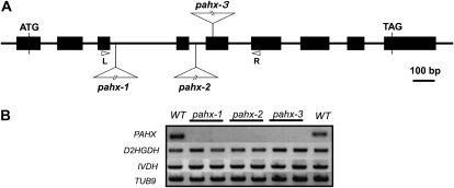
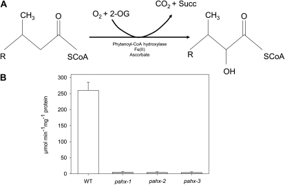
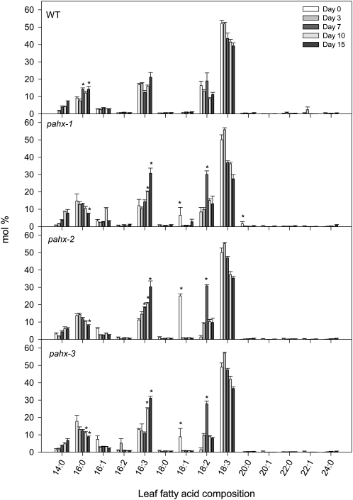
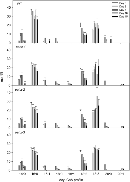
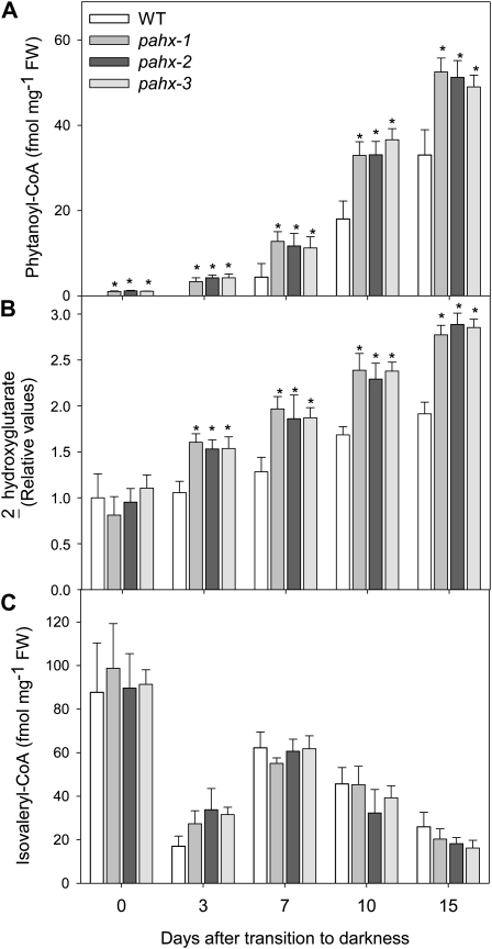
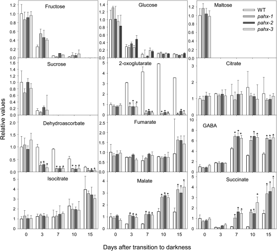
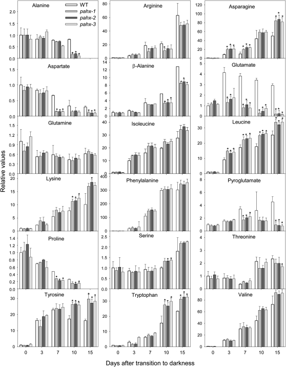
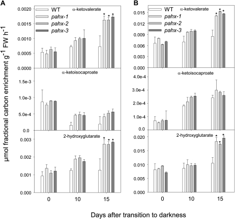
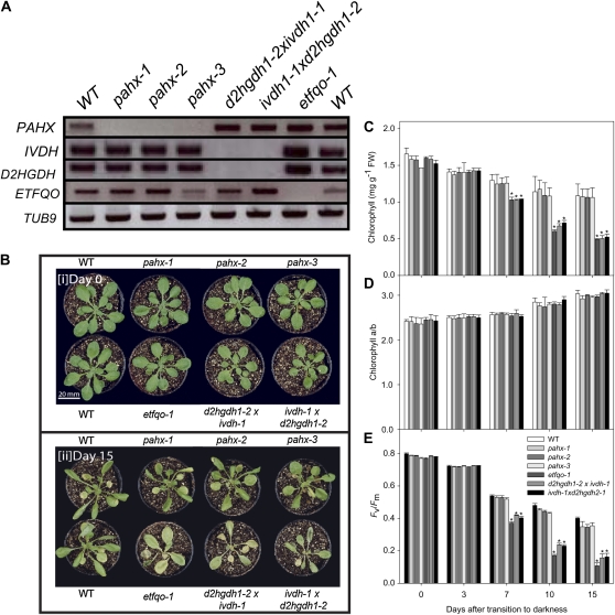
The process of dark-induced senescence in plants is not fully understood, however, the functional involvement of an electron-transfer flavoprotein/electron-transfer flavoprotein:ubiquinone oxidoreductase (ETF/ETFQO), has been demonstrated. Recent studies have revealed that the enzymes isovaleryl-coenzyme A (CoA) dehydrogenase and 2-hydroxyglutarate dehydrogenase act as important electron donors to this complex. In addition both enzymes play a role in the breakdown of cellular carbon storage reserves with isovaleryl-CoA dehydrogenase being involved in degradation of the branched-chain amino acids, phytol, and lysine while 2-hydroxyglutarate dehydrogenase is exclusively involved in lysine degradation. Given that the chlorophyll breakdown intermediate phytanoyl-CoA accumulates dramatically both in knockout mutants of the ETF/ETFQO complex and of isovaleryl-CoA dehydrogenase following growth in extended dark periods we have investigated the direct importance of chlorophyll breakdown for the supply of carbon and electrons during this process. For this purpose we isolated three independent Arabidopsis (Arabidopsis thaliana) knockout mutants of phytanoyl-CoA 2-hydroxylase and grew them under the same extended darkness regime as previously used. Despite the fact that these mutants accumulated phytanoyl-CoA and also 2-hydroxyglutarate they exhibited no morphological changes in comparison to the other mutants previously characterized. These results are consistent with a single entry point of phytol breakdown into the ETF/ETFQO system and furthermore suggest that phytol is not primarily metabolized by this pathway. Furthermore analysis of isovaleryl-CoA dehydrogenase/2-hydroxyglutarate dehydrogenase double mutants generated here suggest that these two enzymes essentially account for the entire electron input via the ETF complex.


Figures

Figure 1.
Figure 1.
A schematic representation of the sites of T-DNA insertion in the pahx mutants. A, Genomic structure of PAHX (At2g01490). Arrowheads represent positions of primers used for genotyping of wild-type and mutant lines; black boxes indicate exons. The T-DNAs are inserted in intron 3, intron 4, and exon 5 of PAHX, in pahx-1, pahx-2, and pahx-3, respectively. B, RT-PCR analysis on total RNA (two biological replicates) from the wild type (WT) and the pahx-1, pahx-2, and pahx-3 mutant lines, with primer sets for the genes indicated on the left. The positions of the primers (R and L) in the PAHX genomic locus are represented in A.
Figure 2.
Figure 2.
Conversion of phytanoyl-CoA to α-hydroxyphytanoyl-CoA by the Fe(II)- and 2-oxoglutarate-dependent enzyme phytanoyl-coenzyme A α-hydroxylase (PAHX), with coevolution of carbon dioxide and succinic acid. A, Schematic reaction catalyzed by PAHX and enzyme activity of pahx mutant lines compared to the wild-type (WT) control (Col 0; B).The activity was determined in leaves of 4-week-old, short-day-grown, Arabidopsis plants. Values are means ± se of five independent samplings.
Figure 3.
Figure 3.
Leaf fatty acid composition in pahx Arabidopsis mutants plants during growth under extended dark conditions. Data (in % mol) represent means ± se for six independent samplings of the ninth or 10th leaves of 4-week-old, short-day-grown, Arabidopsis plants after further treatment for 0, 3, 7, 10, and 15 d in extended darkness. Fatty acid composition was analyzed by GC of fatty acid methyl esters. An asterisk indicates values that were determined by the Student’s t test to be significantly different (P < 0.05) from the wild type at the same time point.
Figure 4.
Figure 4.
Acyl-CoA profiles in pahx Arabidopsis mutants plants during growth under extended dark conditions. Data (in % mol) represent means ± se for six independent samplings of the ninth or 10th leaves of 4-week-old, short-day-grown, Arabidopsis plants after further growth for 0, 3, 7, 10, and 15 d in extended darkness. Acyl-CoAs in samples of 10 mg (fresh weight) each were derivatized to their acyl-etheno-CoA esters, separated by HPLC, and detected fluorometrically.
Figure 5.
Figure 5.
Metabolic phenotype of pahx Arabidopsis mutants plants during growth under extended dark conditions. Phytanoyl-CoA (A), 2-hydroxyglutarate (B), and isovaleryl-CoA (C) profiles in pahx Arabidopsis mutants under extended dark treatment. Samples were taken from the ninth or 10th leaves of 4-week-old, short-day-grown, Arabidopsis plants after further growth for 0, 3, 7, 10, and 15 d in extended darkness. Values are means ± se of six independent samplings; an asterisk indicates values that were determined by the Student’s t test to be significantly different (P < 0.05) from the wild type.
Figure 6.
Figure 6.
Relative levels of sugars and organic acids in Arabidopsis mutant plants during growth under extended dark conditions as measured by GC-MS. The y axis values represent the metabolite level relative to wild type. Data were normalized to the mean response calculated for the 0-d dark-treated leaves of the wild type (in case no response was detected at 0 d, normalization was performed against 3-d dark-treated leaves of the wild type). Values presented are means ± se of determinations on six independent samplings; an asterisk indicates values that were determined by the Student’s t test to be significantly different (P < 0.05) from the wild type.
Figure 7.
Figure 7.
Relative levels of amino acids in Arabidopsis mutants plants during growth under extended dark conditions as measured by GC-MS. Levels of the indicated amino acids are presented as in Figure 6.
Figure 8.
Figure 8.
Redistribution of 13C label following incubation of Arabidopsis mutants and wild-type leaves. The incubation was followed via the transpiration stream in [U-13C] Val (A) or [U-13C] Lys (B) solution. The leaf material was harvested from the ninth or 10th leaves of 4-week-old, short-day-grown, Arabidopsis plants after growth for 0, 10, and 15 d in extended darkness. Values represent absolute redistribution and are given as means ± se of determinations on six independent samplings; an asterisk indicates values that were determined by the Student’s t test to be significantly different (P < 0.05) from the wild type.
Figure 9.
Figure 9.
Phenotype of ETF/ETFQO-related Arabidopsis mutants during growth under extended dark conditions. A, RT-PCR analysis on total RNA from the wild type (WT) and the pahx-1, pahx-2, and pahx-3 mutant lines, as well as two double knockout lines ivdhh1-1 × d2hgdh1-2 and etfqo-1 with primer sets for the genes indicated on the left. B, Images of 4-week-old, short-day-grown Arabidopsis plants immediately (0 d) and after further growth for up to 15 d in darkness conditions. The leaves of the pahx mutant lines were only partially yellowed and dehydrated following 15 d growth in darkness compared to the wild-type (WT) control (Col0). Chlorophyll content (C), chlorophyll a/b ratio (D), and Fv/Fm (E), the maximum quantum yield of PSII electron transport, of leaves of 4-week-old, short-day-grown, Arabidopsis plants after further growth for 0, 3, 7, 10, and 15 d in extended darkness. Values are means ± se of six independent samplings; an asterisk indicates values that were determined by the Student’s t test to be significantly different from the wild type (P < 0.05).

References

    1. Araújo WL, Ishizaki K, Nunes-Nesi A, Larson TR, Tohge T, Krahnert I, Witt S, Obata T, Schauer N, Graham IA, et al. (2010) Identification of the 2-hydroxyglutarate and isovaleryl-CoA dehydrogenases as alternative electron donors linking lysine catabolism to the electron transport chain of Arabidopsis mitochondria. Plant Cell 22: 1549–1563 - PMC - PubMed
    1. Araújo WL, Tohge T, Ishizaki K, Leaver CJ, Fernie AR. (2011) Protein degradation—an alternative respiratory substrate for stressed plants. Trends Plant Sci (in press) - PubMed
    1. Beckmann JD, Frerman FE. (1985) Electron-transfer flavoprotein-ubiquinone oxidoreductase from pig liver: purification and molecular, redox, and catalytic properties. Biochemistry 24: 3913–3921 - PubMed
    1. Bradford MM. (1976) A rapid and sensitive method for the quantitation of microgram quantities of protein utilizing the principle of protein-dye binding. Anal Biochem 72: 248–254 - PubMed
    1. Bräutigam A, Weber AP. (2011) Do metabolite transport processes limit photosynthesis? Plant Physiol 155: 43–48 - PMC - PubMed

Publication types