

Review
doi: 10.1083/jcb.201011152.
Evolution: Tracing the origins of centrioles, cilia, and flagella
Affiliations
- PMID: 21788366
- PMCID: PMC3144413
- DOI: 10.1083/jcb.201011152
Item in Clipboard
Review
Evolution: Tracing the origins of centrioles, cilia, and flagella
J Cell Biol.
.
Erratum in
- J Cell Biol. 2011 Oct 17;195(2):341
Abstract
Centrioles/basal bodies (CBBs) are microtubule-based cylindrical organelles that nucleate the formation of centrosomes, cilia, and flagella. CBBs, cilia, and flagella are ancestral structures; they are present in all major eukaryotic groups. Despite the conservation of their core structure, there is variability in their architecture, function, and biogenesis. Recent genomic and functional studies have provided insight into the evolution of the structure and function of these organelles.
Figures
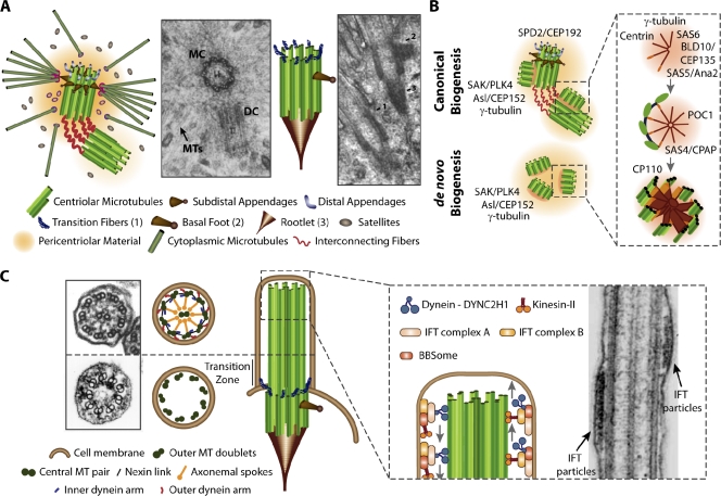
Structure and biogenesis of centrosomes and cilia. (A) On the left, a schematic and EM micrograph (reproduced from Vorobjev and Chentsov, 1982) of an animal prometaphase centrosome composed of mother (MC) and daughter (DC) centriole arranged in an orthogonal fashion. The mother centriole harbors subdistal and distal appendages. On the right, a schematic and EM longitudinal section (reproduced with permission from the Journal of Cell Science; Sorokin, 1968) of a basal body from rat lung multiciliated cells bearing rootlets and lateral/distal appendages. (B) Key regulatory and structural components in CBB biogenesis (canonical [top] and de novo [bottom]; Azimzadeh and Marshall, 2010). (C) Schematic of the basal body, when docked at the cell membrane and growing the axoneme of cilia/flagella. EM cross section of tracheal motile cilia (top: reproduced from Satir and Dirksen (1985) in Handbook of Physiology with permission from the American Physiology Association) and renal nonmotile primary cilia (bottom: image courtesy of H. Zentgraf, German Cancer Research Center, Heidelberg, Germany). Cilia/flagella are assembled via the intraflagellar transport (IFT) system. EM longitudinal section of the Chlamydomonas flagellum adapted from Pedersen et al. (2006) with permission from Elsevier.
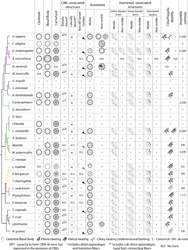
Structure and distribution of CBB, cilia/flagella, and associated structures in eukaryotes. Simplified taxonomic tree representing major eukaryotic groups in different colors (these groups contain a common ancestor and all its descendants; adapted from Hedges (2002) and Baldauf (2003)). Unikonts include eukaryotic cells that, for the most part, have a single emergent flagellum and are divided into Opisthokonts (propel themselves with a single posterior flagellum; Metazoans, Fungi, and Choanoflagellates) and Amoebozoa (Cavalier-Smith, 2002). Bikonts include eukaryotic organisms with two emergent flagella (Cavalier-Smith, 2002). Branch color code: purple, Opisthokonts; blue, Amoebozoa; green, Plants; yellow, Alveolates; orange, Stramenopiles; rose, Rhizaria; brown, Excavates and Discicristates. We represent the symmetry and number of CBB MTs, either when nucleating an axoneme (basal body) or not (centriole), and of axonemes as well as the presence/absence of central MT pair (−, not present). In organisms for which data were available, we also included the structure of the cartwheel and of several associated structures (transition fibers/distal appendages involved in CBB anchoring at the cell membrane; lateral appendages including subdistal appendages, basal foot or connecting fibers linking CBBs to each other or to cytoskeleton components; and non-MT based rootlets that link the proximal part of the CBB to other organelles). We also included information on the presence/absence of axonemal-associated structures (dynein arms and radial spokes) as well as their beating patterns. Finally, we represent the pathways used for CBB assembly, canonical, de novo, or both. In Drosophila melanogaster, the gray centriolar MTs represent the fact that certain tissues present centrioles with doublets whereas others show triplets. The asterisk in D. melanogaster 9+0 sensory axoneme reflects the possibility that this structure is motile (Göpfert and Robert, 2003). In Acerentomon microrhinus, the CBBs formed during spermatogenesis have a different symmetry and the cartwheel is also represented in these structures. In the remaining cases, gray structures are used when evidence pointing to their presence is not robust (poor EM data or data from a related species). For references please check Table S1 .
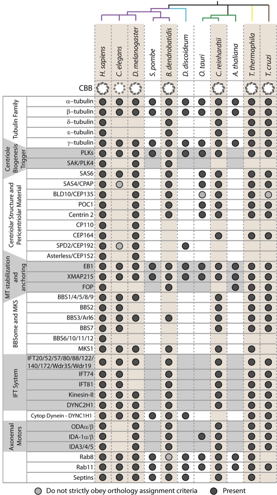
Phylogenetic profile of CBB/axoneme structure and components. Simplified taxonomic tree representing major eukaryotic groups in different colors using the same color code as in Fig. 2 (adapted from Hedges (2002) and Baldauf (2003)). Phylogenetic profile of proteins involved in CBB and cilia/flagella assembly and function. Data adapted from Jékely and Arendt (2006), Wickstead and Gull (2007), Carvalho-Santos et al. (2010), Hodges et al. (2010), Wickstead et al. (2010) and our unpublished data.
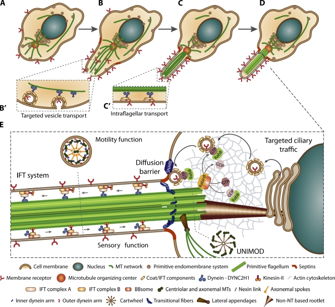
Autogenous theory for the origin of the sensory/motile flagellum. (A) The eukaryotic cell where the proto-cilium evolved likely had a cytoskeleton composed of actin and MTs that converged in the MTOC, a nucleus, and an endomembrane system. (B and B′) Targeted traffic of cell membrane components to a cell membrane patch that started protruding through the directed force produced by the MTs anchored at the MTOC. This protrusion would evolve to become a specialized structure with specific membrane composition maintained by diffusion barriers. (C) The evolving proto-cilium was likely capable of environmental sensing and gliding, which might have driven the implementation of this organelle. (C′) An IFT system was recruited to assemble these structures. (D) Further on, the bundle of MTs would evolve in order to create a specialized arrangement of closed and open MTs forming a ninefold symmetric structure capable of bending due to the presence of molecular motors. The basal body gave support to the motile axoneme at the cell membrane. (E) In conclusion: the ancestral CBB/cilium apparatus would have been characterized by the following characteristics: (a) a ninefold symmetric CBB composed of MT triplets, with a cartwheel, lateral and distal appendages, and rootlets being defined by a set of proteins, the UNIMOD (UNIversal MODule); (b) an axoneme with both motile and sensorial functions presenting ninefold symmetry composed of nine doublets, central pair, outer and inner dynein arms, and maintained by the IFT system; (c) a specialized membrane created by a diffusion barrier both at the level of the membrane and of diffusion of components at the transition zone; and (d) targeted transport of membrane and other components from the Golgi to the ciliary base. Adapted from Satir et al. (2008).
References
-
- Azimzadeh J., Bornens M. 2004. The centrosome in evolution. Centrosomes in Development and Disease. Nigg E.A., editor Wiley-VCH, Weinheim: 93–122
Publication types
MeSH terms
LinkOut - more resources
Full Text Sources
