. 2011 Sep 1;187(5):2213-21.
doi: 10.4049/jimmunol.1003952. Epub 2011 Jul 25.

Silencing microRNA-155 ameliorates experimental autoimmune encephalomyelitis

Affiliations

Silencing microRNA-155 ameliorates experimental autoimmune encephalomyelitis

Gopal Murugaiyan et al. J Immunol. .

Abstract

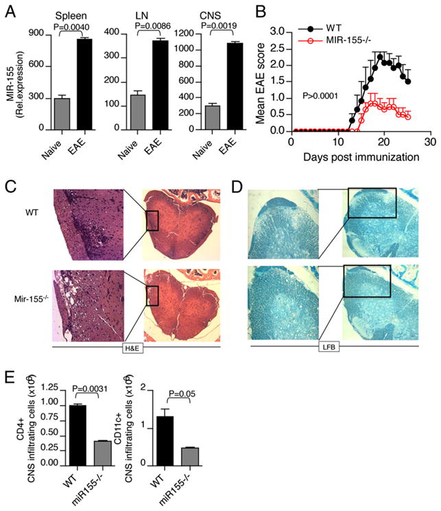
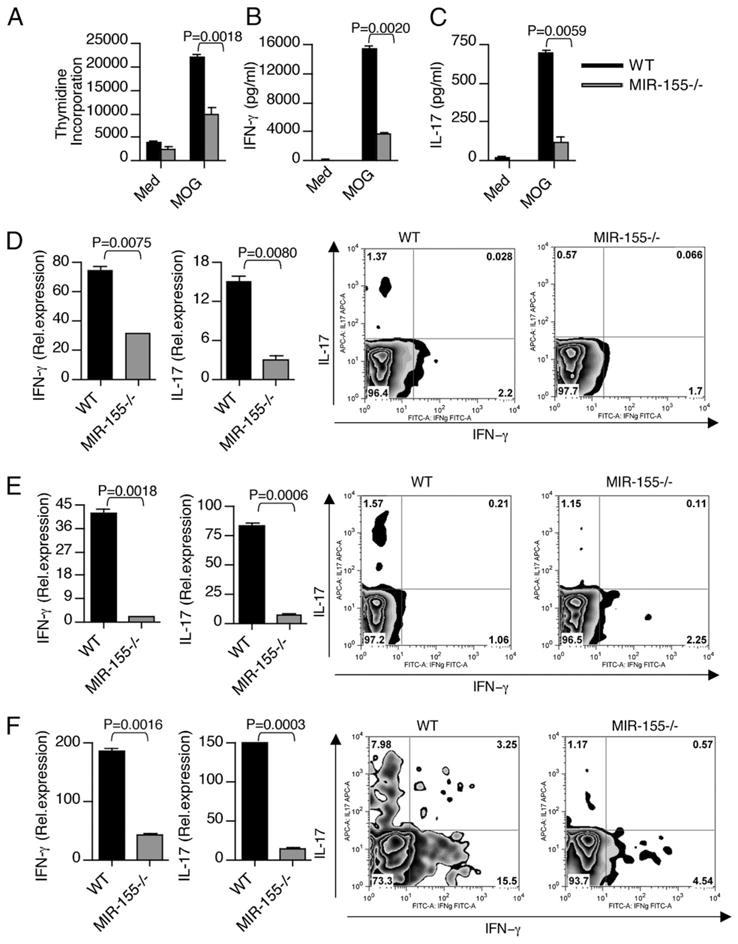
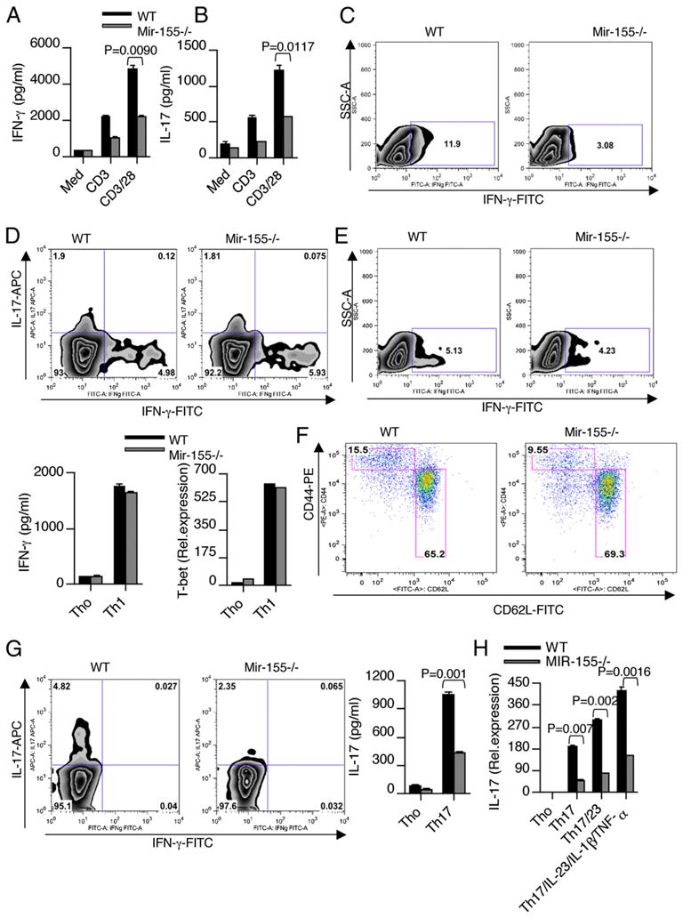
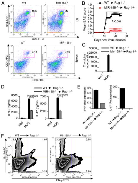
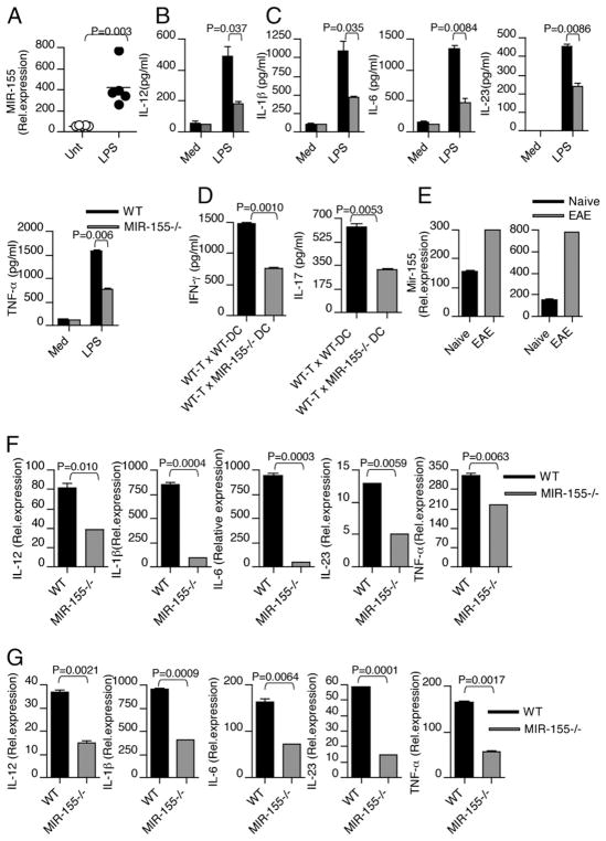
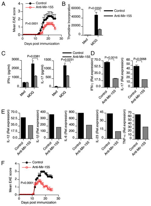
IFN-γ-producing Th1 and IL-17-producing Th17 cells are the key participants in various autoimmune diseases, including multiple sclerosis and its animal model, experimental autoimmune encephalomyelitis (EAE). Although both of these T cell subsets are known to be regulated by specific transcription factors and cytokines, the role of microRNAs that control these two inflammatory T cell subsets and whether targeting microRNAs can have therapeutic effects are not known. In this study, we show that microRNA-155 (Mir-155) expression is elevated in CD4(+) T cells during EAE, and Mir-155(-/-) mice had a delayed course and reduced severity of disease and less inflammation in the CNS. The attenuation of EAE in Mir-155(-/-) mice was associated with a decrease in Th1 and Th17 responses in the CNS and peripheral lymphoid organs. The T cell-intrinsic function of Mir-155(-/-) was demonstrated by the resistance of Mir-155(-/-) CD4(+) T cell-repleted Rag-1(-/-) mice to EAE. Finally, we found that anti-Mir-155 treatment reduced clinical severity of EAE when given before and after the appearance of clinical symptoms. These findings demonstrate that Mir-155 confers susceptibility to EAE by affecting inflammatory T cell responses and identify Mir-155 as a new target for therapeutic intervention in multiple sclerosis.


Conflict of interest statement

Disclosures

The authors have no financial conflicts of interest.

Figures

FIGURE 1
FIGURE 1
Mir-155−/− mice develop attenuated EAE. Elevated Mir-155 expression in CD4+ T cells during EAE. A, Mir-155 mRNA was determined by quantitative real-time PCR analysis in CD4+ T cells from spleen, LN, and CNS from naive and EAE mice. B, Mir-155−/− mice develop attenuated EAE. Clinical scores of WT mice (WT; n = 8) and Mir-155−/− mice (Mir-155−/−; n = 8) at various times after immunization with MOG(33–55) in CFA. Results are representative of three independent experiments. C and D, Histopathological analysis of spinal cord sections from representative of WT and Mir-155−/− mice (n = 3/group) at 20 d after immunization. Spinal cord sections were stained with H&E and luxol fast blue to access inflammation and demyelination. Original magnification ×20 (C) and ×40 (D). E, Number of CNS-infiltrating CD4+ T cells and CD11c+ cells. Mononuclear cells were isolated from spinal cords of WT and Mir-155−/− mice (n = 3/group) at 20 d after immunization, and then they were pooled and counted. Cells were stained with Abs specific for cell surface markers, as indicated, and the percentage of each subset is shown.
FIGURE 2
FIGURE 2
Mir-155−/− mice exhibit lower Th1 and Th17 cytokine production during EAE. Splenocytes from WT and Mir-155−/− mice were harvested 10 d after immunization and were stimulated ex vivo with MOG peptide 35–55. A, In the last 16 h, cells were pulsed with thymidine and assayed for proliferation (cpm). Error bars represent SEM between triplicates. Supernatants from parallel cultures were harvested 72 h after initiation of cultures and assayed by ELISA for IFN-γ (B) and IL-17 (C). Real-time RT-PCR analysis and intracellular staining of IFN-γ and IL-17 in (D) spleen-, (E) LN-, and (F) CNS-derived CD4+ T cells isolated from WT and Mir-155−/− mice with EAE. Data in A–F are representative of three experiments.
FIGURE 3
FIGURE 3
Mir-155 regulates CD4+ T cell effector cytokine production. Total CD4+ T cells were isolated from the spleens of WT and Mir-155−/− mice and stimulated with anti-CD3 and anti-CD28. Supernatants from cultures were harvested 72 h after initiation of cultures and analyzed for cytokines (A, B) IFN-γ and IL-17. Intracellular staining was used to determine the number of IFN-γ–producing cells (C). Data are representative of three independent experiments. D, Naive CD4+ T cells (CD4+CD62LhighCD44low) from spleens were purified by flow cytometry and stimulated with plate-bound Ab against CD3 (2 μg/ml) and CD28 (2 μg/ml) under Th1-skewing condition. Intracellular staining was used to determine the number of Th1 cells. Supernatants from cultures were harvested 4 d after initiation of cultures and analyzed for cytokines IFN-γ by ELISA. Similarly, stimulated cells were harvested 48 h poststimulation for mRNA and analyzed for T-bet by quantitative real-time PCR. Data are representative of five independent experiments. E, Memory CD4+ T cells (CD4+CD62LlowCD44high) from spleens were purified by flow cytometry and stimulated with plate-bound Ab against CD3 (2 μg/ml) and CD28 (2 μg/ml). Intracellular staining was used to determine the number of IFN-γ–producing cells. Data are representative of three independent experiments. F, Flow cytometry analysis of CD44high memory CD4+ T cells in the spleens of WT and Mir-155−/− mice. Naive CD4+ T cells (CD4+CD62LhighCD44low) from spleens were purified by flow cytometry and stimulated with plate-bound Ab against CD3 (2 μg/ml) and CD28 (2 μg/ml) under Th17-skewing condition. G, Intracellular staining was used to determine the number of Th17 cells. H, IL-17 mRNA was determined by quantitative real-time PCR analysis. Data are representative of three to five independent experiments.
FIGURE 4
FIGURE 4
T cell-intrinsic effect of Mir-155 in EAE. Rag-1−/− mice (n = 5/group) were injected with either WT or Mir-155−/− CD4+ T cells. Seven d after transfer, mice were harvested, and engraftment of CD3+CD4+ T cells was assayed by flow cytometry with splenocytes and LN (A). Rag-1−/− mice (n = 5/group) were injected with either WT or Mir-155−/− CD4+ T cells. Seven d after transfer, mice were immunized with MOG peptide emulsified in CFA and were monitored for EAE (B). Results are representative of two independent experiments. C, Splenocytes from these mice were activated in vitro with MOG p35–55 (20 μg/ml). Proliferation was measured by [3H]thymidine incorporation assay. Error bars represent SEM between triplicates. D, Cytokines were measured in culture supernatants using ELISA. Data are representative of two experiments. E and F, Real-time RT-PCR and flow cytometry analysis of IFN-γ and IL-17 in CNS-derived CD4+ T cells isolated from Rag-1−/− mice reconstituted with T cells from WT and Mir-155−/− mice.
FIGURE 5
FIGURE 5
Mir-155 positively regulates Th1-and Th17-polarizing cytokine expression in DCs. Bone marrow-derived DCs isolated from WT mice (n = 5/group) were stimulated with LPS. A, Mir-155 mRNA was determined by quantitative real-time PCR analysis in DCs in response to LPS. B and C, Bone marrow-derived DCs were isolated from WT and Mir-155−/− mice and were cultured with 100 ng/ml LPS. Cell-free supernatants were measured for Th1- and Th17-polarizing cytokine by ELISA. D, CD4+ T cells isolated from WT mice were cocultured with CD11c+ DCs derived from WT and Mir-155−/− mice. Supernatants from cultures were harvested 72 h after initiation of cultures and assayed by ELISA for IFN-γ and IL-17. E, Mir-155 mRNA was determined by quantitative real-time PCR analysis in DCs isolated from LN and CNS of mice with and without EAE (n = 4/group). Data are representative of two independent experiments. Real-time PCR measurement of Th1-and Th17-polarizing cytokines in DCs isolated from spleen (F) and LN (G) of WT and Mir-155−/− mice with EAE. Data are representative of three experiments.
FIGURE 6
FIGURE 6
Anti–Mir-155 treatment reduces the clinical severity of EAE. A, Clinical scores of EAE in WT mice (n = 5/group) treated with anti–Mir-155 or scrambled control on days 5, 7, 9, and 11 postimmunization. Results are representative of two independent experiments. B, Splenocytes obtained from anti–Mir-155 and control mice were restimulated with MOG35–55 (20 μg/ml) for 72 h. Proliferation was measured by [3H]thymidine incorporation assay. Error bars represent SEM between triplicates. C, Cell-free supernatants were assayed for IL-17 and IFN-γ by ELISA. D, Real-time RT-PCR analysis of IFN-γ and IL-17 in CNS-derived CD4+ T cells isolated from mice treated with or without anti–Mir-155. E, Real-time PCR measurement of Th1- and Th17-polarizing cytokines in DCs isolated from LN of control and anti–Mir-155–treated mice (n = 5/group). Data are representative of two independent experiments. F, Mice treated with Mir-155 inhibitor after onset of EAE (score of ≥1.5) resulted in a rapid clinical recovery from EAE.

References

    1. Diveu C, McGeachy MJ, Cua DJ. Cytokines that regulate autoimmunity. Curr Opin Immunol. 2008;20:663–668. - PubMed
    1. Windhagen A, Newcombe J, Dangond F, Strand C, Woodroofe MN, Cuzner ML, Hafler DA. Expression of costimulatory molecules B7-1 (CD80), B7-2 (CD86), and interleukin 12 cytokine in multiple sclerosis lesions. J Exp Med. 1995;182:1985–1996. - PMC - PubMed
    1. Lock C, Hermans G, Pedotti R, Brendolan A, Schadt E, Garren H, Langer-Gould A, Strober S, Cannella B, Allard J, et al. Gene-microarray analysis of multiple sclerosis lesions yields new targets validated in autoimmune encephalomyelitis. Nat Med. 2002;8:500–508. - PubMed
    1. Kebir H, Ifergan I, Alvarez JI, Bernard M, Poirier J, Arbour N, Duquette P, Prat A. Preferential recruitment of interferon-gamma-expressing TH17 cells in multiple sclerosis. Ann Neurol. 2009;66:390–402. - PubMed
    1. Langrish CL, Chen Y, Blumenschein WM, Mattson J, Basham B, Sedgwick JD, McClanahan T, Kastelein RA, Cua DJ. IL-23 drives a pathogenic T cell population that induces autoimmune inflammation. J Exp Med. 2005;201:233–240. - PMC - PubMed

Publication types

MeSH terms