. 2011 Aug 30;108(35):14431-6.
doi: 10.1073/pnas.1104221108. Epub 2011 Jul 25.

Flexibility of the C-terminal, or CII, ring of KaiC governs the rhythm of the circadian clock of cyanobacteria

Affiliations

Flexibility of the C-terminal, or CII, ring of KaiC governs the rhythm of the circadian clock of cyanobacteria

Yong-Gang Chang et al. Proc Natl Acad Sci U S A. .

Abstract

In the cyanobacterial circadian oscillator, KaiA and KaiB alternately stimulate autophosphorylation and autodephosphorylation of KaiC with a periodicity of approximately 24 h. KaiA activates autophosphorylation by selectively capturing the A loops of KaiC in their exposed positions. The A loops and sites of phosphorylation, residues S431 and T432, are located in the CII ring of KaiC. We find that the flexibility of the CII ring governs the rhythm of KaiC autophosphorylation and autodephosphorylation and is an example of dynamics-driven protein allostery. KaiA-induced autophosphorylation requires flexibility of the CII ring. In contrast, rigidity is required for KaiC-KaiB binding, which induces a conformational change in KaiB that enables it to sequester KaiA by binding to KaiA's linker. Autophosphorylation of the S431 residues around the CII ring stabilizes the CII ring, making it rigid. In contrast, autophosphorylation of the T432 residues offsets phospho-S431-induced rigidity to some extent. In the presence of KaiA and KaiB, the dynamic states of the CII ring of KaiC executes the following circadian rhythm: CII STflexible → CIISpTflexible → CIIpSpTrigid → CIIpSTvery-rigid → CIISTflexible. Apparently, these dynamic states govern the pattern of phosphorylation, ST → SpT → pSpT → pST → ST. CII-CI ring-on-ring stacking is observed when the CII ring is rigid, suggesting a mechanism through which the ATPase activity of the CI ring is rhythmically controlled. SasA, a circadian clock-output protein, binds to the CI ring. Thus, rhythmic ring stacking may also control clock-output pathways.


Conflict of interest statement

The authors declare no conflict of interest.

Figures

Fig. 1.
Fig. 1.
Phosphorylation kinetics profiles of KaiC and KaiC phosphomimics. Each data point represents the average of two experiments. The error bars represent the SEM. (A) Phosphorylation of KaiC in the presence and/or absence of KaiA and KaiB. KaiC alone (red circle); KaiC+KaiA (dark blue diamond); KaiC+KaiB (green triangle); KaiC+KaiA+KaiB (purple square). (B) Phosphorylation of KaiC variants KaiC-AT (dark blue diamond), KaiC-SE (green triangle), KaiC-SA (purple cross), and KaiC-ET (cyan square) in the presence of KaiA. These profiles were obtained by densitometric analysis of SDS-PAGE gels (SI Appendix, Fig. S1). Phosphorylation profiles of these KaiC variants in the absence of KaiA are shown in SI Appendix, Fig. S1.
Fig. 2.
Fig. 2.
CII ring flexibility depends on the state of phosphorylation at residues S431 and T432 and A-loop positions. (AC) Selected regions from methyl-TROSY (54) spectra of U-[15N,2H]-Ile-δ1-[13C,1H]-labeled KaiC (panel 1) and phosphomimics of SpT-KaiC (panel 2), pSpT-KaiC (panel 3), pST-KaiC (panel 4), and pSpT-KaiC487 (panel 5). Horizontal and vertical axes are 1H and 13C chemical shifts in ppm, respectively. All panels were plotted at the same contour level relative to the signal (SI Appendix, Table S1). Gel-filtration experiments indicated that these KaiC mutants did not aggregate. Red and blue contours indicate peaks assigned to Ile residues of the CII and CI domains of KaiC, respectively, whereas black contours indicate unassigned peaks. See Fig. S2A for full spectra, Fig. S2B for peak assignments, Table S1 for plotting parameters, and Table S3 for NMR experimental details in SI Appendix. It should be noted that KaiC-SE is approximately 30% phosphorylated at residue S431 (see SI Appendix, Fig. S1C). (D) Gel-filtration profiles of isolated domains of CII (panel 1) and phosphomimics of SpT-CII (panel 2), pSpT-CII (panel 3), pST-CII (panel 4), and pSpT-CII487 and pST-CII487 (panel 5). The hexameric forms of the CII domains eluted at approximately 11.7 mL, whereas the monomeric forms eluted at approximately 15.4 mL. In panel 5, CII-EE487 and CII-ET487, two KaiC CII domains that were truncated just prior to the A loops at residues 488, eluted as monomers at approximately 16 mL. The percentages of hexameric (H) and monomeric forms (M) are provided in each panel.
Fig. 3.
Fig. 3.
KaiC-KaiB binding experiments. (A) Gel-filtration profiles of mixtures of KaiB with KaiC (panel 1) and with phosphomimics of SpT-KaiC (panel 2), pSpT-KaiC (panel 3), pST-KaiC (panel 4), and pSpT-KaiC487 (panel 5). The elution profiles of KaiB in each panel are from the same run. (B) Methyl-TROSY spectra of the U-[15N,2H]-Ile-δ1-[13C,1H]-labeled pSpT-KaiC phosphomimic free (black contours) and bound (red contours) to unlabeled KaiB. The two spectra were plotted at the same contour level. (C) Methyl-TROSY spectra of U-[15N,2H]-Ile-δ1-[13C,1H]-labeled KaiB free (black contours) and bound (red contours) to the unlabeled pSpT-KaiC phosphomimic. “x”s indicate artifact peaks. Please see SI Appendix, Table S3 for experimental details. The two spectra were plotted at the same contour level.
Fig. 4.
Fig. 4.
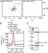
Mapping the KaiCB binding site on KaiA. (A) Titration of KaiA-130C (residues 130–283) with KaiC-EE497 and KaiB. (Left) Free KaiA-130C; (Right) KaiA-130C + KaiC-EE497 + KaiB. The monomeric molar ratio of KaiA-130C, KaiC-EE497, and KaiB was 2∶3∶2. Sample conditions and NMR parameters are described in SI Appendix, Table S3. The two spectra were plotted at the same contour level. (B) Gel-filtration chromatography showing the coelution of KaiA-130C, KaiB, and KaiC-EE497. The NMR sample for A, Right was loaded onto a Superdex 200 10/300 GL column to determine whether the absence of NMR signals was due to sample aggregation. Arrows indicate peaks for free KaiC-EE497 (red peak) and the complex formed by KaiA-130C, KaiB, and KaiC-EE497 (black peak). The apparent molecular size of the complex (approximately 440 kDa) was close to the size expected for a complex composed of a KaiC-EE497 hexamer, two KaiA-130C dimers, and two KaiB dimers (460 kDa) (27, 55). (C) SDS-PAGE gel of the elution peak indicated by the black arrow in B. Protein bands in lane 2 corresponding to KaiC-EE497, KaiA-130C, and KaiB are indicated. Molecular weight markers are in lane 1.
Fig. 5.
Fig. 5.
CII-CI interactions depend on the state of phosphorylation at residues S431 and T432. (A) Gel-filtration experiments of mixtures of domains of CI with CII (panel 1) and phosphomimics of SpT-CII (panel 2), pSpT-CII (panel 3), pST-CII (panel 4), and pST-CII487 (panel 5). The hexameric form of the CI and CII domains eluted at approximately 11.7 mL, whereas the monomeric forms eluted at approximately 15.4 mL. The shaded region in panel 4 indicates the shift of the CI + pST-CII phosphomimic elution profile relative to those of the domains by themselves. (B) Selected regions of methyl-TROSY spectra of U-[15N,2H]-Ile-δ1-[13C,1H]-labeled (29) KaiC (panel 1) and the phosphomimics of SpT-KaiC (panel 2), pSpT-KaiC (panel 3), pST-KaiC (panel 4), and pSpT-KaiC-487 (panel 5). Horizontal and vertical axes are 1H and 13C chemical shifts in ppm, respectively. All panels were plotted at the same relative contour level. Blue resonances are those assigned to the CI domain, whereas unassigned peaks are black. Corresponding regions of methyl-TROSY spectra of free CI are shown in panel 6.

Comment in

  • Dynamic fluctuations lubricate the circadian clock.
    Pai MT, Kalodimos C. Pai MT, et al. Proc Natl Acad Sci U S A. 2011 Aug 30;108(35):14377-8. doi: 10.1073/pnas.1111105108. Epub 2011 Aug 23. Proc Natl Acad Sci U S A. 2011. PMID: 21873208 Free PMC article. No abstract available.

References

    1. Albrecht U, editor. The Circadian Clock. New York: Springer; 2010.
    1. Dunlap JC, Loros JJ, DeCoursey PJ, editors. Chronobiology: Biological Timekeeping. Sunderland, MA: Sinauer; 2004.
    1. Ditty JL, Mackey SR, Johnson CH, editors. Bacterial Circadian Programs. Heidelberg: Springer; 2009.
    1. Ishiura M, et al. Expression of a gene cluster kaiABC as a circadian feedback process in cyanobacteria. Science. 1998;281:1519–1523. - PubMed
    1. Iwasaki H, Nishiwaki T, Kitayama Y, Nakajima M, Kondo T. KaiA-stimulated KaiC phosphorylation in circadian timing loops in cyanobacteria. Proc Natl Acad Sci USA. 2002;99:15788–15793. - PMC - PubMed

Publication types

MeSH terms

Substances

LinkOut - more resources