. 2011 Aug 9;108(32):13194-9.
doi: 10.1073/pnas.1018456108. Epub 2011 Jul 25.

Craniofacial divergence and ongoing adaptation via the hedgehog pathway

Affiliations

Craniofacial divergence and ongoing adaptation via the hedgehog pathway

Reade B Roberts et al. Proc Natl Acad Sci U S A. .

Abstract

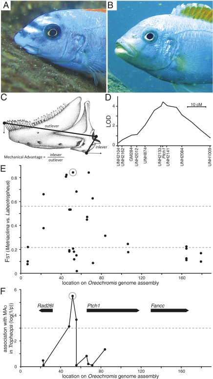
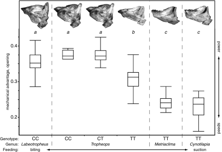
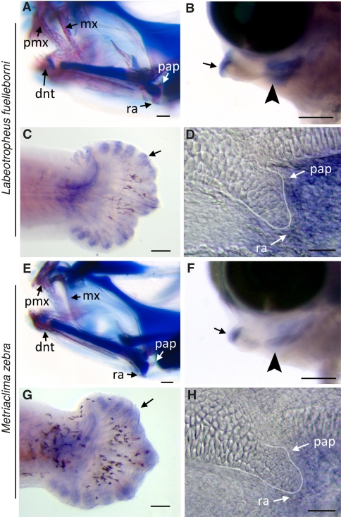
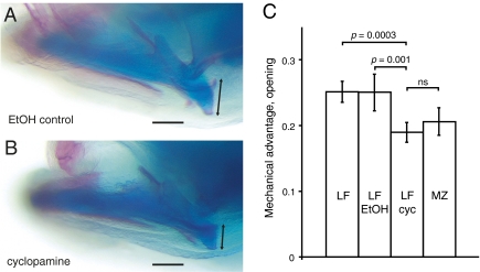
Adaptive variation in craniofacial structure contributes to resource specialization and speciation, but the genetic loci that underlie craniofacial adaptation remain unknown. Here we show that alleles of the hedgehog pathway receptor Patched1 (Ptch1) gene are responsible for adaptive variation in the shape of the lower jaw both within and among genera of Lake Malawi cichlid fish. The evolutionarily derived allele of Ptch1 reduces the length of the retroarticular (RA) process of the lower jaw, a change predicted to increase speed of jaw rotation for improved suction-feeding. The alternate allele is associated with a longer RA and a more robustly mineralized jaw, typical of species that use a biting mode of feeding. Genera with the most divergent feeding morphologies are nearly fixed for different Ptch1 alleles, whereas species with intermediate morphologies still segregate variation at Ptch1. Thus, the same alleles that help to define macroevolutionary divergence among genera also contribute to microevolutionary fine-tuning of adaptive traits within some species. Variability of craniofacial morphology mediated by Ptch1 polymorphism has likely contributed to niche partitioning and ecological speciation of these fishes.


Conflict of interest statement

The authors declare no conflict of interest.

Figures

Fig. 1.
Fig. 1.
Ptch1 locus characterization in families, genera, and populations. Craniofacial appearance of (A) Labeotropheus trewavasae (image courtesy of Justin Marshall) and (B) Metriaclima mbenjii. (C) Lower jaw phenotype measures; RA and inlever length is equivalent. (D) QTL mapping interval for MAO on cichlid LG12. (E) Population differentiation (FST) between Labeotropheus and Metriaclima (n = 24 each) for SNPs at the Ptch1 locus. Dashed lines indicate two experimental measures of mean FST; the lower line from global comparison of Labeotropheus vs. Metriaclima across many populations (20), and the upper line from comparisons of randomized Labeotropheus and Metriaclima population pairs from distinct sites (48). (F) Significance of association between SNPs at the Ptch1 locus and MAO in the genus Tropheops (Wald test, n = 59); dashed line indicates a P value of 0.001. Genes in the region annotated with bold arrows. The SNP used to indicate long and short alleles of Ptch1 is circled in E and F.
Fig. 2.
Fig. 2.
Morphology and function of the lower jaw by genus and Ptch1 genotype. MAO values of wild individuals (Table S2) indicated by box-plot, with letters above designating statistical groups by one-way ANOVA and Tukey's honest significance test (P < 0.05). Lower jaw images from representative individuals at top; each jaw has an MAO matching the mean of the corresponding genus and genotype. Genotype refers to high-association SNP (circled in Fig. 1 E and F). Box boundaries indicate quartiles, and whiskers indicate range of MAO data.
Fig. 3.
Fig. 3.
Interspecific differences in Ptch1 expression during cichlid craniofacial development. Craniofacial outcomes in (A) LF and (E) MZ larvae at 13 dpf; LF larvae exhibit accelerated bone (pink) development compared with MZ; cartilage stains blue. Ptch1 in situ labeling in representative 6-dpf (stage 12) (B–D) LF and (F–H) MZ larvae: (B and F) Lateral whole-mount view of lower jaw with nodes of Ptch1 expression, particularly in mesenchymal cells at the distal (arrow) and proximal (arrowhead) ends of the lower jaw precursor where the dentary and RA process will form, respectively. (C and G) Ptch1 labeling is qualitatively similar in developing fin-ray elements of tail (arrows). (D and H) Flat-mount preparation of the jaw joint in lateral view, showing the cartilaginous precursor of the RA process (outlined) relative to node of Ptch1 expression. dnt, dentary; mx, maxilla; pap, posterior articulation process; pmx, premaxilla. (Scale bars, 200 μm in A, B, E, and F; 100 μm in C and G; and 10 μm in D and H.)
Fig. 4.
Fig. 4.
Treatment of biting species larvae with a hedgehog pathway inhibitor recapitulates a suction-feeding jaw phenotype. LF larvae were treated with (A) 0.5% ethanol (EtOH control, n = 7) or (B) 50 μM cyclopamine (cyc, n = 10) for 6 h at stage 12; RA length measured at 12 dpf (black arrow in A and B). (C) Relative to the length of the lower jaw (measured as the distance between the center of the jaw joint and the tip of the dentary), RA length (measured as the distance from the posterior tip of the posterior articulation process to the ventral tip of the retroarticular) was significantly reduced in cyclopamine treatment group compared with the control LF group but was not significantly different from MZ control larvae (n = 7). Larvae treated with ethanol were indistinguishable from untreated siblings (n = 6). P values, one-way ANOVA. (Scale bar, 100 μm.)

References

    1. Gans C, Northcutt RG. Neural crest and the origin of vertebrates: A new head. Science. 1983;220:268–273. - PubMed
    1. Darwin CR. Journal of Researches into the Geology and Natural History of the Various Countries Visited During the Voyage of H.M.S. Beagle, Under the Command of Captain FitzRoy, R.N. 2nd Ed. London: John Murray; 1845.
    1. Grant PR, Grant BR. Unpredictable evolution in a 30-year study of Darwin's finches. Science. 2002;296:707–711. - PubMed
    1. Abzhanov A, Protas M, Grant BR, Grant PR, Tabin CJ. Bmp4 and morphological variation of beaks in Darwin's finches. Science. 2004;305:1462–1465. - PubMed
    1. Abzhanov A, et al. The calmodulin pathway and evolution of elongated beak morphology in Darwin's finches. Nature. 2006;442:563–567. - PubMed

MeSH terms

Associated data

LinkOut - more resources