. 2011 Aug 23;108(34):14306-11.
doi: 10.1073/pnas.1101767108. Epub 2011 Jul 25.

A diversity of paracrine signals sustains molecular circadian cycling in suprachiasmatic nucleus circuits

Affiliations

A diversity of paracrine signals sustains molecular circadian cycling in suprachiasmatic nucleus circuits

Elizabeth S Maywood et al. Proc Natl Acad Sci U S A. .

Abstract

The suprachiasmatic nucleus (SCN) is the principal circadian pacemaker of mammals, coordinating daily rhythms of behavior and metabolism. Circadian timekeeping in SCN neurons revolves around transcriptional/posttranslational feedback loops, in which Period (Per) and Cryptochrome (Cry) genes are negatively regulated by their protein products. Recent studies have revealed, however, that these "core loops" also rely upon cytosolic and circuit-level properties for sustained oscillation. To characterize interneuronal signals responsible for robust pacemaking in SCN cells and circuits, we have developed a unique coculture technique using wild-type (WT) "graft" SCN to drive pacemaking (reported by PER2::LUCIFERASE bioluminescence) in "host" SCN deficient either in elements of neuropeptidergic signaling or in elements of the core feedback loop. We demonstrate that paracrine signaling is sufficient to restore cellular synchrony and amplitude of pacemaking in SCN circuits lacking vasoactive intestinal peptide (VIP). By using grafts with mutant circadian periods we show that pacemaking in the host SCN is specified by the genotype of the graft, confirming graft-derived factors as determinants of the host rhythm. By combining pharmacological with genetic manipulations, we show that a hierarchy of neuropeptidergic signals underpins this paracrine regulation, with a preeminent role for VIP augmented by contributions from arginine vasopressin (AVP) and gastrin-releasing peptide (GRP). Finally, we show that interneuronal signaling is sufficiently powerful to maintain circadian pacemaking in arrhythmic Cry-null SCN, deficient in essential elements of the transcriptional negative feedback loops. Thus, a hierarchy of paracrine neuropeptidergic signals determines cell- and circuit-level circadian pacemaking in the SCN.


Conflict of interest statement

The authors declare no conflict of interest.

Figures

Fig. 1.
Fig. 1.
Restoration of SCN circadian gene expression by coculture. (A) Representative bioluminescence rhythm from VIP-null SCN grafted with nonreporter WT SCN after 10 d. Note damping followed by immediate restoration of rhythms by graft. (B) Relative amplitude (percentage of initial peak value) of successive peaks of circadian bioluminescence of VIP-null SCN recorded for 10 d and then given grafts of SCN, PVN, or cerebral cortex (plotted as mean ± SEM, n ≥ 5). (C) Period of bioluminescence rhythm of VIP-null SCN (mean ± SEM) grafted with WT, Tau, or Afh mutant SCN (n ≥ 4). **P = 0.01 vs. WT treatment (ANOVA, F = 163.8). (D) Representative raster plot of circadian gene expression in 50 cells from WT SCN with accompanying Raleigh plot. (E and F) Representative raster plot from VIP-null SCN before (E) and after (F) grafting with WT SCN. Progressive loss of synchrony and its restoration are reflected in Rayleigh plots on days −9, −1, and +3 relative to grafting.
Fig. 2.
Fig. 2.
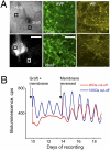
Paracrine signaling of circadian cues between SCN. (A) Confocal reconstruction of representative VIP-null SCN with WT graft. (Left) Low power (10×) phase and bioluminescence images identifying boxed areas examined at higher power. (Scale bar, 1 mm.) (Right) Confocal images (60×) for AVP- and VIP-ir in graft and host tissues. (Scale bar, 50 μm.) (B) Representative bioluminescence rhythm from VIP-null SCN grafted after 10 d with WT SCN separated by 2-kDa (red) or 10-kDa (blue) cutoff membranes. After 3 d the grafts were placed directly on the host.
Fig. 3.
Fig. 3.
Restoration of SCN circadian gene expression by signals other than VIP. (A) Representative bioluminescence emission from VPAC2-null SCN grafted with nonreporter WT SCN after 10 d. Note damping of rhythm before grafting, followed by progressive restoration of rhythm. (B) Relative amplitude (percentage of initial peak value) of successive peaks of circadian bioluminescence of WT SCN (green, n = 4) recorded for 20 d (mean ± SEM) or of VPAC2-null (red, n = 6) or VIP-null (blue, n = 6) SCN recorded for 10 d and then given grafts of WT SCN. (C) Representative raster plot of circadian bioluminescence in 50 cells from VPAC2-null SCN with accompanying Raleigh plots from days −9, −1, +3, and +6 relative to grafting. Note progressive loss of synchrony and its restoration.
Fig. 4.
Fig. 4.
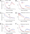
Absence of VIP/VPAC2 signaling reveals dependence of SCN circadian gene expression on AVP- and GRP-mediated signaling. (A) Relative amplitude (percentage of initial peak value) of successive peaks of circadian bioluminescence of VIP-null SCN grafted with WT SCN and treated with vehicle (n = 3) or GRP receptor antagonist (n = 4, mean ± SEM). (B) As in A but with AVP V1a and V1b antagonists (vehicle, n = 4; antagonists, n = 5). (C) Representative bioluminescence recordings of VPAC2-null SCN grafted with WT SCN and treated with vehicle or GRP antagonist. (D) As in C but treatment with vehicle or AVP V1a and V1b receptor antagonists. (E) Relative amplitude (percentage of initial peak value) of successive peaks of circadian bioluminescence of VPAC2-null SCN grafted with WT SCN and treated with vehicle (n = 5) or GRP receptor blocker (n = 6, mean ± SEM). (F) As in E but with vehicle (n = 5) or AVP V1a and V1b receptor antagonists (n = 7).
Fig. 5.
Fig. 5.
Residual circadian pacemaking in Cry-null SCN slices and cells and its coordination by interneuronal signaling. (A) Representative coherent circadian bioluminescence rhythms of Cry-null SCN. (B) Aggregate bioluminescence emission of representative nonrhythmic Cry-null SCN and individual raster plots from 50 cells. (C) Representative bioluminescence emission from (nonrhythmic) Cry-null (black) and VIP-null (blue) SCN grafted with WT SCN. Note immediate restoration of rhythms in VIP-null and progressive effect in Cry-null SCN. (D) Raster and Rayleigh plots (with accompanying mean vector) of bioluminescence of 50 cells in representative Cry-null SCN that received a WT graft.

Comment in

  • Neuropeptides go the distance for circadian synchrony.
    Freeman GM Jr, Herzog ED. Freeman GM Jr, et al. Proc Natl Acad Sci U S A. 2011 Aug 23;108(34):13883-4. doi: 10.1073/pnas.1110844108. Epub 2011 Aug 11. Proc Natl Acad Sci U S A. 2011. PMID: 21836054 Free PMC article. No abstract available.

References

    1. Takahashi JS, Hong HK, Ko CH, McDearmon EL. The genetics of mammalian circadian order and disorder: Implications for physiology and disease. Nat Rev Genet. 2008;9:764–775. - PMC - PubMed
    1. Meng QJ, et al. Setting clock speed in mammals: The CK1 epsilon tau mutation in mice accelerates circadian pacemakers by selectively destabilizing PERIOD proteins. Neuron. 2008;58:78–88. - PMC - PubMed
    1. Godinho SI, et al. The after-hours mutant reveals a role for Fbxl3 in determining mammalian circadian period. Science. 2007;316:897–900. - PubMed
    1. Hastings MH, Maywood ES, O'Neill JS. Cellular circadian pacemaking and the role of cytosolic rhythms. Curr Biol. 2008;18:R805–R815. - PubMed
    1. Mohawk JA, Takahashi JS. Cell autonomy and synchrony of suprachiasmatic nucleus circadian oscillators. Trends Neurosci. 2011;34:349–358. - PMC - PubMed

Publication types

MeSH terms

Substances