. 2011 Nov;91(11):1668-79.
doi: 10.1038/labinvest.2011.113. Epub 2011 Jul 25.

P-glycoprotein induction by breast milk attenuates intestinal inflammation in experimental necrotizing enterocolitis

Affiliations

P-glycoprotein induction by breast milk attenuates intestinal inflammation in experimental necrotizing enterocolitis

Yigit S Guner et al. Lab Invest. 2011 Nov.

Abstract

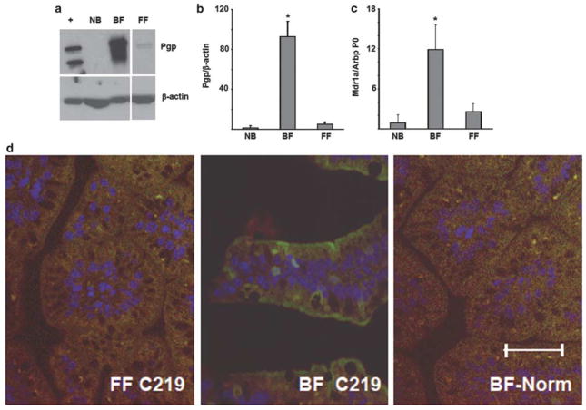
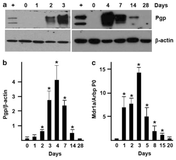
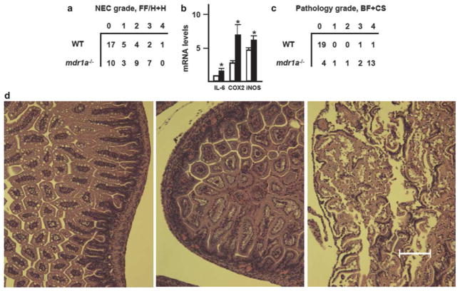
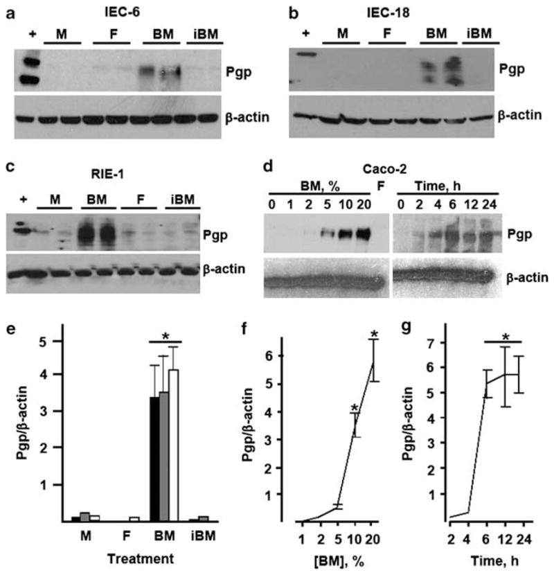
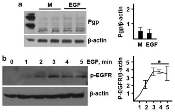
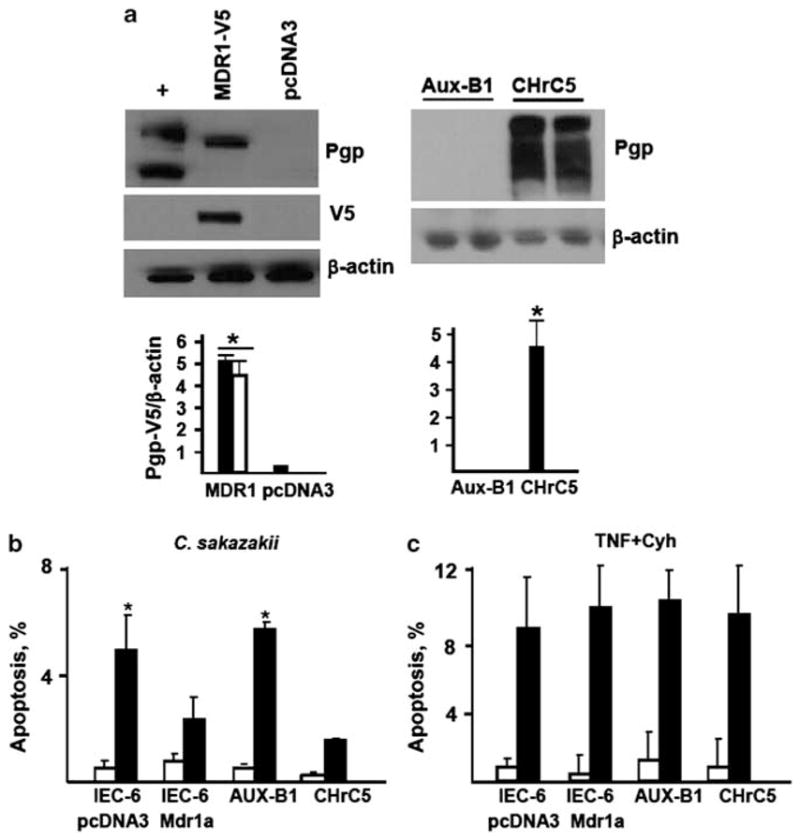
P-glycoprotein (Pgp), a product of the multi-drug resistance gene MDR1a, is a broad specificity efflux ATP cassette transmembrane transporter that is predominantly expressed in epithelial tissues. Because mdr1a(-/-) mice tend to develop spontaneous colitis in bacteria-dependent manner, Pgp is believed to have a role in protection of the intestinal epithelium from luminal bacteria. Here we demonstrate that levels of Pgp in the small intestine of newborn rodents dramatically increase during breastfeeding, but not during formula feeding (FF). In rats and mice, levels of intestinal Pgp peak on days 3-7 and 1-5 of breastfeeding, respectively. The mdr1a(-/-) neonatal mice subjected to FF, hypoxia, and hypothermia have significantly higher incidence and pathology, as well as significantly earlier onset of necrotizing enterocolitis (NEC) than congenic wild type mice. Breast-fed mdr1a(-/-) neonatal mice are also more susceptible to intestinal damage caused by the opportunistic pathogen Cronobacter sakazakii that has been associated with hospital outbreaks of NEC. Breast milk, but not formula, induces Pgp expression in enterocyte cell lines in a dose- and time-dependent manner. High levels of ectopically expressed Pgp protect epithelial cells in vitro from apoptosis induced by C. sakazakii. Taken together, these results show that breast milk-induced expression of Pgp may have a role in the protection of the neonatal intestinal epithelium from injury associated with nascent bacterial colonization.


Conflict of interest statement

DISCLOSURE/CONFLICT OF INTEREST

The authors declare no conflict of interest.

Figures

Figure 1
Figure 1
Breastfeeding increases expression of P-glycoprotein (Pgp) in the small intestine. (a) Western blots for Pgp protein levels in mucosal scrapings from terminal ileum of newborn (NB) rats immediately after birth, breast-fed (BF) for 4 days, and subjected to formula feeding/hypoxia (FF/H) for 4 days. β-actin re-probes are shown to demonstrate lane load. ‘ +’, positive control (lysate of CHrC5 cells). Pgp electromobility variations are presumably due to different degrees of glycosylation. Images are representative of five independent experiments. (b) Average Pgp protein band densities. Pgp levels were normalized to those of β-actin in the same sample. *Significant difference from NB or FF (P<0.05). (c) Average levels of Mdr1a mRNA in the terminal ileum of BF and FF/H rats as determined by real-time RT-PCR. Levels of Mdr1a mRNA were normalized to those of Arbp P0 mRNA in the same sample. *Significant difference from NB or FF (P<0.05). (d) Small intestinal sections from 4-day-old breast-fed (BF) or formula-fed (FF) rats immunostained with C219 antibody (Ab), or normal mouse serum as indicated. Green, Pgp immunofluorescence; blue, DAPI-stained nuclei. Bar = 100 μM. Images are representative of three independent experiments.
Figure 2
Figure 2
Timing of P-glycoprotein (Pgp) expression in the terminal ileum of breast-fed (BF) rodents. (a) A representative western blot for Pgp and β-actin protein levels in mucosal scrapings from terminal ileum of BF rats at the indicated age. ‘ +’, positive control (CHrC5 lysate). (b) Average Pgp protein band densities. Pgp levels were normalized to those of β-actin in the same sample. *Significant difference from day 0 (n = 4, P<0.05). (c) Average levels of Mdr1a mRNA in small intestinal mucosa of FVB mice on the indicated days of breastfeeding. Levels of Mdr1a mRNA were normalized to those of Arbp P0 mRNA in the same sample. *Significant difference from day 0 (n = 3, P<0.05).
Figure 3
Figure 3
Mdr1a deficiency predisposes mice to necrotizing enterocolitis (NEC). (a) Pathology grades in wild-type FVB and mdr1a−/− mice subjected to the FF/H +H regimen. Incidence and grades of NEC are significantly higher in the mdr1a−/− group (n = 29, P<0.01, χ2-test; P<0.05, two-tailed Mann–Whitney test, respectively). (b) Expression of IL-6, cyclooxygenase-2 (COX-2), and inducible nitric oxide synthase (iNOS) mRNAs in the small intestinal mucosa of wild-type (open bars) and mdr1a−/− (filled bars) mice on days 2–3 of the FF/H + H regimen. mRNA levels are relative to Arbp P0 transcript levels, which were assigned the value of 1. *Significant differences from wild type (n = 5, P<0.05). (c) Pathology grades in breast-fed (BF) wild-type FVB and mdr1a−/− mice, challenged with 105 cfu C. sakazakii on day 1. Samples of small intestine were collected on day 4. Pathology grades are significantly higher in the mdr1a−/− group (P = 0.02, n = 21, two-tailed Mann–Whitney test). (d) Representative pathology micrographs. Left, normal architecture, absence of pathology, grade 0; middle, epithelial sloughing, and partial destruction of villus tips, grade 2; right, submucosal edema, and severe disruption of villus architecture, grade 3. Bar = 100 μM.
Figure 4
Figure 4
Breast milk induces P-glycoprotein (Pgp) in cultured enterocytes. (ac) IEC-6, IEC-18, or RIE-1 cells were treated for 24 h with medium (M), 5% centrifugation-cleared formula (F), 5% cleared breast milk (BM), or 5% cleared breast milk incubated at 60° C for 10 min (iBM), as indicated. Levels of Pgp and β-actin were examined by western blotting. For each cell line and treatment, two independently treated samples are shown. (d) Dose response (left) and time course (right) of Pgp expression in Caco-2 cells treated with human breast milk for 24 h at indicated concentrations (left), or 10% human breast milk for indicated time. ‘ +’, positive control (CHrC5 cells). Data are representative of at least three independent experiments. (e) Average Pgp band densities on western blots represented by (ac). *Significant differences from other treatment groups (P<0.01, n = 3). (f) Dose response of Pgp induction in Caco-2 cells by human breast milk. *Significant difference from other timepoints (P<0.05, n = 3 for each concentration). (g) Time course of breast milk-induced expression of Pgp in Caco-2 cells. *Significant differences from other time points (P<0.05, n = 3 for each time point).
Figure 5
Figure 5
Epidermal growth factor (EGF) does not induce P-glycoprotein (Pgp) in IEC-6 cells. (a) Pgp and β-actin levels in IEC-6 cells treated with medium (M) or 100 ng/ml EGF, as indicated, for 24 h. ‘ +’, Positive control (CHrC5 cells). (b) Phospho-EGFR and β-actin levels in IEC-6 cells treated with 100 ng/ml EGF for indicated time. *Significantly higher phospho-EGFR levels compared with other time points (P<0.05, n = 3).
Figure 6
Figure 6
P-glycoprotein (Pgp) protects epithelial cells from C. sakazakii-induced apoptosis. (a) Expression of V5-tagged Pgp protein in IEC-6 cells stably transfected with pcDNA3-MDR1-V5 or pcDNA3 analyzed by Western blotting with Pgp and V5 epitope antibody (Abs; left); expression of Pgp protein in CHrC5 and AuxB1 cells (right). β-actin blots are shown to demonstrate lane load. (+), positive control (CHrC5 cells). Solid bars, Pgp immunoreactivity; open bars, V5 immunoreactivity. *Significant differences from cells transfected with pcDNA3 (P<0.05, n = 3) or from Aux-B1 cells (P<0.01, n = 3). (b) Average frequencies of spontaneous (open bars) and C. sakazakii-induced (filled bars) apoptosis in indicated cell lines. *Significant differences compared to the cell line expressing Pgp (n = 3, P<0.05). (c) Average frequencies of spontaneous (open bars) or tumor necrosis factor (TNF-α) + cycloheximide-induced apoptosis in indicated cell lines.

References

    1. Guthrie SO, Gordon PV, Thomas V, et al. Necrotizing enterocolitis among neonates in the United States. J Perinatol. 2003;23:278–285. - PubMed
    1. Hammerman C, Bin-Nun A, Kaplan M. Germ warfare: probiotics in defense of the premature gut. Clin Perinatol. 2004;31:489–500. - PubMed
    1. Morowitz MJ, Poroyko V, Caplan M, et al. Redefining the role of intestinal microbes in the pathogenesis of necrotizing enterocolitis. Pediatrics. 125:777–785. - PubMed
    1. Patole S. Prevention and treatment of necrotising enterocolitis in preterm neonates. Early Hum Dev. 2007;83:635–642. - PubMed
    1. Hall NJ, Eaton S, Peters MJ, et al. Mild controlled hypothermia in preterm neonates with advanced necrotizing enterocolitis. Pediatrics. 2010;125:e300–e308. - PubMed

Publication types

MeSH terms

Substances