. 2011;6(7):e21799.
doi: 10.1371/journal.pone.0021799. Epub 2011 Jul 18.

IL-22 is produced by innate lymphoid cells and limits inflammation in allergic airway disease

Affiliations

IL-22 is produced by innate lymphoid cells and limits inflammation in allergic airway disease

Christian Taube et al. PLoS One. 2011.

Abstract

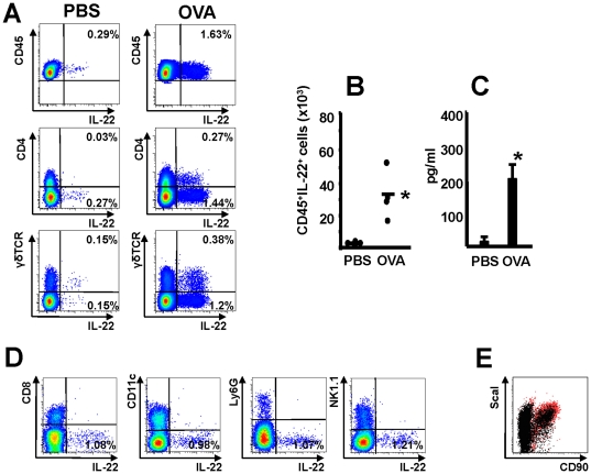
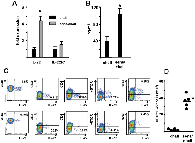
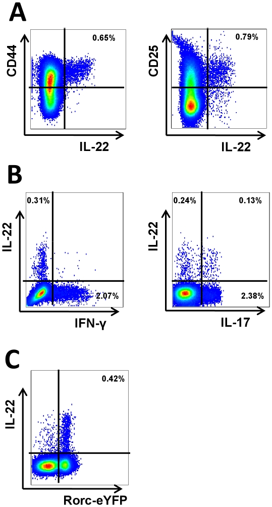
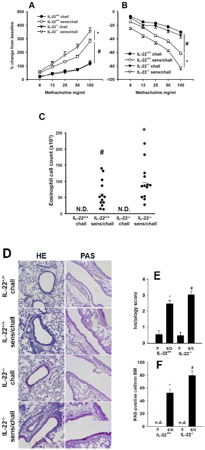
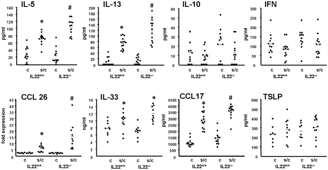
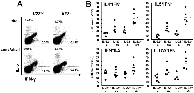
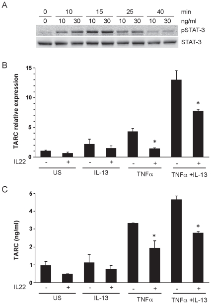
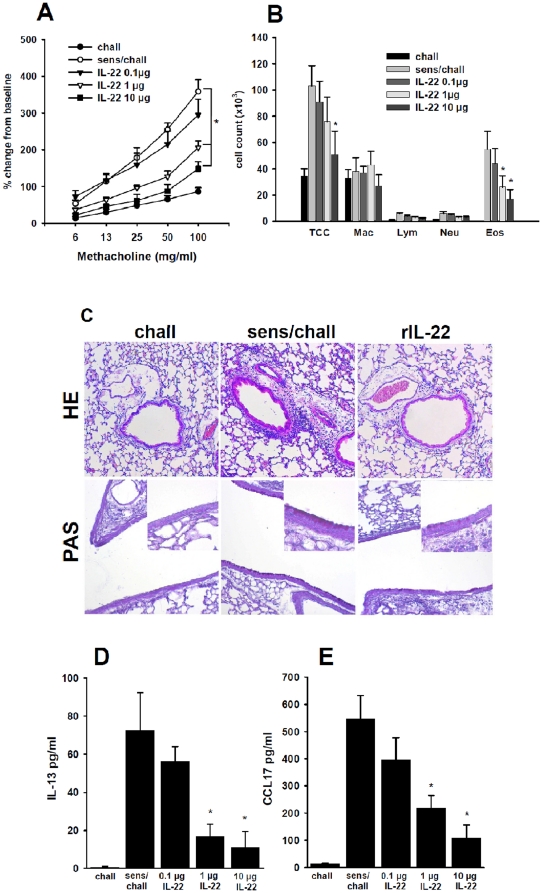
Interleukin (IL)-22 is an effector cytokine, which acts primarily on epithelial cells in the skin, gut, liver and lung. Both pro- and anti-inflammatory properties have been reported for IL-22 depending on the tissue and disease model. In a murine model of allergic airway inflammation, we found that IL-22 is predominantly produced by innate lymphoid cells in the inflamed lungs, rather than TH cells. To determine the impact of IL-22 on airway inflammation, we used allergen-sensitized IL-22-deficient mice and found that they suffer from significantly higher airway hyperreactivity upon airway challenge. IL-22-deficiency led to increased eosinophil infiltration lymphocyte invasion and production of CCL17 (TARC), IL-5 and IL-13 in the lung. Mice treated with IL-22 before antigen challenge displayed reduced expression of CCL17 and IL-13 and significant amelioration of airway constriction and inflammation. We conclude that innate IL-22 limits airway inflammation, tissue damage and clinical decline in allergic lung disease.


Conflict of interest statement

Competing Interests: The authors have declared that no competing interests exist.

Figures

Figure 1
Figure 1. IL-22 expression is increased during specific T cell responses in the lung.
Panel A: IL-22 intracellular staining in lung cells 24 hrs after inhaled exposure of OT II mice with either PBS (PBS) or OVA for 3 consecutive days. Panel A shows IL-22 a representative intracellular staining of CD45+ cells. Panel B: Numbers of CD45+ IL22+ cells from 2 independent experiments, each dot represents a single mouse, bar represents Mean, * p<0.05; Panel C: levels of IL-22 in BAL fluid, mean±SEM are shown, n = 4 from 2 independent experiments, Panel D: representative IL-22 intracellular staining in lung cells 24 hrs after inhaled exposure of OT II mice OVA for 3 consecutive days. Panel E: ScaI and CD90 expression in lung cells 24 hrs after inhaled exposure of OT II mice. Red dots represent IL-22 positive cells, black dots represent IL-22 negative cells.
Figure 2
Figure 2. IL-22 expression is increased during allergic airway inflammation.
Panel A: Expression of IL-22 and IL-22 R1 (IL-22 Rc) was assessed in lung tissue of challenged only (chall, n = 6) and sensitized and challenged (sens/chall, n = 6) animals. Total RNA was isolated 24 hours after the last challenge, reverse transcribed, and gene expression analyzed by PCR with specific primers for IL-22R1. Data are shown as fold induction relative to expression in naïve animals after normalization to GAPDH. Mean±SEM from 2 independent experiments are given. * p<0.05. Panel B: Levels of IL-22 in BAL fluid 48 hrs following the last challenge in challenged only (chall, n = 6) and sensitized and challenged (sens/chall, n = 6) animal. Mean±SEM from 2 independent experiments are given. * p<0.05. Panel C and D: IL-22 intracellular staining in lung cells 24 hrs following the last exposure in sensitized and challenged (top row) and challenged only (bottom row) animals and frequency of CD45+IL-22+ cells in lung tissue each dot represents a single mouse from 2 independent experiments. * p<0.05.
Figure 3
Figure 3. Analysis of cytokine production and surface markers of infiltrating mononuclear cells in the lungs.
Panel A shows the expression of CD25 and CD44 among IL-22 producing cells. Panel B shows the IFN-γ and IL-17A production from sensitized and challenged mouse lungs. Panel C shows the expression of Rorgt. Rorc-eYFP mice were sensitized and challenged and the lung infiltrating mononuclear cells were analyzed for the expression of YFP.
Figure 4
Figure 4. IL-22 deficient animals display increased AHR and airway inflammation.
Airway resistance (panel A) and dynamic compliance (panel B) in challenged only wild-type (IL-22+/+ chall, n = 12), challenged only IL-22 deficient (IL-22−/− chall, n = 12), sensitized and challenged wild-type (IL-22+/+ sens/chall, n = 13) and sensitized and challenged IL-22 deficient (IL-22−/− sens/chall, n = 13) mice. Results are expressed as mean±SEM from 2 independent experiments. # p<0.01, *p<0.05. Panel C shows differential cell counts for eosinophils in BAL fluid. Each dot represents a single mouse, bar represents mean. # p<0.05 compared to IL-22+/+ chall and IL-22−/− chall; * p<0.05 compared to all other groups. N.D.: not detectable. Panel D: Tissue inflammation was evaluated 48 hrs following the last challenge using hematoxylin and eosin staining (HE) and PAS staining for goblet cells in challenged only wild-type mice (IL-22+/+ chall), sensitized and challenge wild-type mice (IL-22+/+ sens/chall), challenged only IL-22 deficient mice (IL-22−/− chall) and sensitized and challenged IL-22 deficient (IL-22−/− sens/chall). Final magnifications 100× and 400× for inserts. Panel E shows histology score and panel F number of goblet cells per mm of basement membrane for challenged only (c) and sensitized and challenged (s/c) wild-type (IL-22+/+) and IL-22 deficient (IL-22−/−) animals. Each groups contains 12 animals. Means±SEM are given. *p<0.01 compared to IL-22+/+ c and IL-22−/− c. # p<0.05 compared to all other groups.
Figure 5
Figure 5. Analysis of cytokines and chemokines in IL-22−/− and congenic controls.
Levels of IL-5. IL-13, IL-10, IFN-γ (IFN) and CCL-17 in BAL fluid, TSLP and IL-33 in lung homogenate and expression of CCL26 in whole lung were analyzed in challenged only (c) and sensitized and challenged (s/c) Il22 deficient (IL-22−/− c, n = 12; IL-22−/− s/c, n = 13) and congenic wild-type controls (IL-22+/+ c, n = 12; IL-22+/+ s/c, n = 13). Each dot represents a single mouse, bar represents mean. Data are from 2 independent experiments. * p<0.05 compared to challenged groups, # p<0.05 compared to all other groups.
Figure 6
Figure 6. Intracellular cytokine staining of mononuclear cells in the lung.
Cells were isolated from either challenged only (c) or sensitized and challenged (s/c) Il22 deficient and congenic wild-type controls (IL-22+/+). Panel A shows a representative plot of intracellular IL-5 and IFN-γ staining. Panel B shows cell counts for IL-4 positive, IFN-γ negative (IL-4+IFN), IL-5 positive, IFN-γ negative (IL-5+IFN), IFN-γ positive, IL-5 negative (IFN+IL-5) and IL-17A positive, IFN-γ negative (IL-17A+IFN) cells. Each dot represent a single mouse. Data from 2 independent experiments are given. * p<0.05 compared to all other groups.
Figure 7
Figure 7. IL-22 reduces TNFα/IL-13 induced production of CCL17 in murine Clara cells.
Panel A: C22 cells were stimulated with recombinant IL-22, for the indicated time points. Protein lysates were subjected to SDS page gel electrophoresis and Western blot analysis performed with a pSTAT-3 specific antibody. Detection of total STAT-3 as loading control. Data are representative of 2 independent experiments. Panel B and C: C22 cells were stimulated with recombinant IL-13, TNFα, or a combination of both, for 5 hours or left untreated (US). To assess the effect of IL-22 on TNFα and TNFα/IL-13 induced TARC expression, cells were preincubated with recombinant IL-22 for 4 hours and stimulated with the indicated cytokines for an additional 5 hours in the continued presence of IL-22 (IL-22 +). Controls were incubated with IL-22 only for a total of 9 hours. Relative TARC mRNA expression (panel B) was measured in triplicates by quantitative PCR and normalized to GAPDH expression levels. Results are shown as means±SEM of 5 replicates per treatment pooled from 4 independent experiments. TARC levels in cell culture supernatants (panel C) were assessed by ELISA after 48 hours. Results are shown as mean±SEM of triplicates and of 3 independent experiments. *p<0.01 vs. US, **p<0.001 vs. TNFα alone, ***p<0.05 vs. respective controls stimulated in the absence of IL-22.
Figure 8
Figure 8. Administration of rIL-22 reduces AHR and airway inflammation.
Airway responsiveness (panel A), cell counts in BAL fluid (panel B) and lung tissue inflammation and goblet cell metaplasia (panel C) were assessed in mice 48 h after the last airway challenge. Mice which were sensitized and challenged (sens/chall, n = 12) showed increased airway reactivity and numbers of eosinophils in BAL fluid compared to challenged only mice (chall, n = 5), where no eosinophils were detectable. Intranasal treatment of sensitized and challenge animals with 0.1 µg recombinant IL-22 (IL-22 0.1 µg, n = 12) showed little effects on AHR and inflammation. In contrast, mice treated with either 1 µg (IL-22 1 µg, n = 12) or 10 µg (IL-22 1 µg, n = 12) of recombinant IL-22 showed decreased AHR and number of eosinophils in BAL fluid. Means±SEM are given, *p<0.05 compared to sens/chall. Panel C: Tissue inflammation was evaluated 48 hrs following the last challenge using hematoxylin and eosin staining (HE) and PAS staining for goblet cells in challenged only mice (chall), non-treated sensitized and challenged mice (sens/chall) and sensitized and challenged animals treated with 10 µg of recombinant IL-22 (rIL-22). Final magnifications 100× and 400× for inserts. Panels D and E: Levels of IL-13 (panel D) and CCL17 (panel E) were measured in BAL fluid by ELISA 48 h after the last challenge. Means±SEM of challenged only mice (chall, n = 5), non-treated sensitized and challenged mice (sens/chall, n = 12), and sensitized and challenged mice treated with 0.1 µg (0.1 µg IL-22, n = 12), 1 µg (1 µg IL-22, n = 12) and 10 µg (10 µg IL-22, n = 12) of rIl-22, respectively. Mean±SEM are given. * p<0.05 compared to sens/chall and 0.1 µg IL-22.

References

    1. Dumoutier L, Louahed J, Renauld JC. Cloning and characterization of IL-10-related T cell-derived inducible factor (IL-TIF), a novel cytokine structurally related to IL-10 and inducible by IL-9. J Immunol. 2000;164:1814–1819. - PubMed
    1. Kotenko SV, Izotova LS, Mirochnitchenko OV, Esterova E, Dickensheets H, et al. Identification of the functional interleukin-22 (IL-22) receptor complex: the IL-10R2 chain (IL-10Rbeta) is a common chain of both the IL-10 and IL-22 (IL-10-related T cell-derived inducible factor, IL-TIF) receptor complexes. J Biol Chem. 2001;276:2725–2732. - PubMed
    1. Xie MH, Aggarwal S, Ho WH, Foster J, Zhang Z, et al. Interleukin (IL)-22, a novel human cytokine that signals through the interferon receptor-related proteins CRF2-4 and IL-22R. J Biol Chem. 2000;275:31335–31339. - PubMed
    1. Mosser DM, Zhang X. Interleukin-10: new perspectives on an old cytokine. Immunol Rev. 2008;226:205–218. - PMC - PubMed
    1. Dumoutier L, Van RE, Colau D, Renauld JC. Human interleukin-10-related T cell-derived inducible factor: molecular cloning and functional characterization as an hepatocyte-stimulating factor. Proc Natl Acad Sci U S A. 2000;97:10144–10149. - PMC - PubMed

Publication types

MeSH terms

LinkOut - more resources