. 2011;6(7):e22117.
doi: 10.1371/journal.pone.0022117. Epub 2011 Jul 18.

Gene targeting implicates Cdc42 GTPase in GPVI and non-GPVI mediated platelet filopodia formation, secretion and aggregation

Affiliations

Gene targeting implicates Cdc42 GTPase in GPVI and non-GPVI mediated platelet filopodia formation, secretion and aggregation

Huzoor Akbar et al. PLoS One. 2011.

Abstract

Background: Cdc42 and Rac1, members of the Rho family of small GTPases, play critical roles in actin cytoskeleton regulation. We have shown previously that Rac1 is involved in regulation of platelet secretion and aggregation. However, the role of Cdc42 in platelet activation remains controversial. This study was undertaken to better understand the role of Cdc42 in platelet activation.

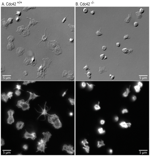
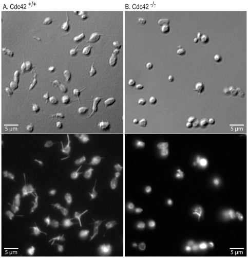
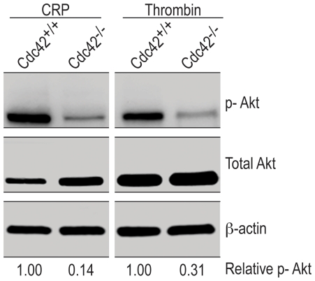
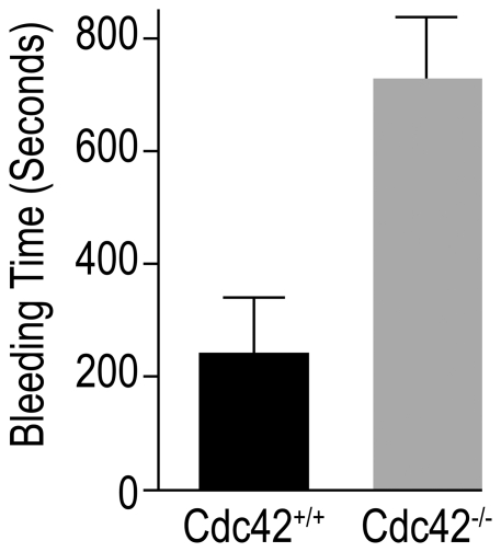
Methodology/principal findings: We utilized the Mx-cre;Cdc42(lox/lox) inducible mice with transient Cdc42 deletion to investigate the involvement of Cdc42 in platelet function. The Cdc42-deficient mice exhibited a significantly reduced platelet count than the matching Cdc42(+/+) mice. Platelets isolated from Cdc42(-/-), as compared to Cdc42(+/+), mice exhibited (a) diminished phosphorylation of PAK1/2, an effector molecule of Cdc42, (b) inhibition of filopodia formation on immobilized CRP or fibrinogen, (c) inhibition of CRP- or thrombin-induced secretion of ATP and release of P-selectin, (d) inhibition of CRP, collagen or thrombin induced platelet aggregation, and (e) minimal phosphorylation of Akt upon stimulation with CRP or thrombin. The bleeding times were significantly prolonged in Cdc42(-/-) mice compared with Cdc42(+/+) mice.

Conclusion/significance: Our data demonstrate that Cdc42 is required for platelet filopodia formation, secretion and aggregation and therefore plays a critical role in platelet mediated hemostasis and thrombosis.


Conflict of interest statement

Competing Interests: The authors have declared that no competing interests exist.

Figures

Figure 1
Figure 1. Gene targeting of Cdc42 induced thrombocytopenia and blocked signaling downstream of Cdc42.
A, Expression of Cdc42, Rac1 and RhoA in platelet lysates from the wild type and genetically targeted mice was probed by Western blotting of Cdc42, Rac1 and RhoA. Platelets from Cdc42 gene targeted mice as compared to the poly (I∶C) treated matching wild type mice showed a complete lack of Cdc42 GTPase. The expression of Rac1 and RhoA was not altered in Cdc42−/− platelets. β-actin expression was used as a loading control. B, Platelet counts (Mean ± SEM) in the Cdc42−/− mice (n = 11) were significantly lower (p<0.05) than the platelet counts in the Cdc42+/+ mice (n = 12). C, CRP (0.2 µg/ml) or thrombin (0.1 U/ml) induced phosphorylation of PAK1/2 is inhibited in the Cdc42−/− mice platelets as compared to the platelets from Cdc42+/+ mice. Phosphorylation of PAK1/2 was analyzed as described in the methods section.
Figure 2
Figure 2. Inducible genetic targeting of Cdc42 inhibited filopodia and lamellipodia formation on immobilized collagen-related peptide.
Washed platelets from Cdc42+/+ (A) and Cdc42−/− (B) mice were layered over cover slips coated with CRP (0.5 µg/ml) for 20 minutes. The cover slips were washed and adherent platelets were processed for DIC (top panel) and immuno-fluorescence (bottom panel) microscopy as detailed in the methods section. Platelets from Cdc42−/− mice failed to form filopodia or lamellipodia on CRP.
Figure 3
Figure 3. Inducible genetic targeting of Cdc42 inhibited filopodia formation on immobilized fibrinogen.
Washed platelets from Cdc42+/+ (A) and Cdc42−/− (B) mice were layered over cover slips coated with fibrinogen (3.0 µg/ml) for 20 minutes. The DIC (top panel) and immuno-fluorescence (bottom panel) images show that platelets from Cdc42−/− mice failed to form filopodia on immobilized fibrinogen.
Figure 4
Figure 4. Inducible genetic targeting of Cdc42 inhibited platelet spreading on immobilized CRP and fibrinogen.
Spreading of washed platelets from Cdc42+/+ and Cdc42−/− mice on CRP or fibrinogen was quantified using Image J software (http://rsbweb.nih.gov/ij). Each bar represents mean surface are ± SEM of 100 platelets. The spreading of Cdc42−/− platelets on immobilized CRP or fibrinogen was significantly (p<0.01) diminished as compared to the Cdc42+/+ platelets.
Figure 5
Figure 5. Genetic targeting of Cdc42 inhibited release of p-selectin from α-granules and secretion of ATP from dense granules.
A, Thrombin (0.2 U/ml) induced expression of P-selectin and B, CRP (0.2 µg/ml) induced secretion of ATP is inhibited in platelets from Cdc42−/− mice compared with the platelets from Cdc42+/+ mice. Bar graphs are the means ± SD (n = 3).
Figure 6
Figure 6. Deficiency of Cdc42 inhibited platelet aggregation induced by collagen, CRP or thrombin.
Washed platelets from Cdc42+/+ and Cdc42−/− were stimulated by addition of CRP (A), collagen (B) or thrombin (C) and aggregation was recorded using a Lumi-Aggregometer at 37°C and a stirring speed of 900 rpm. Platelets from Cdc42−/− mice compared with the platelets from the Cdc42+/+ mice exhibited diminished aggregation induced by all three agonists. The aggregation tracings are representative of three experiments.
Figure 7
Figure 7. Agonist-induced phosphorylation of Akt is reduced in Cdc42−/− platelets.
Washed platelets from Cdc42+/+ and Cdc42−/− mice were incubated with CRP (0.2 µg/ml) or thrombin (0.1 U/ml) for 5 minutes at 37°C with constant stirring in a Chrono-Log aggregometer. The reactions were terminated by adding 5× sample buffer, processed for Western blotting and probed for Akt, p-Akt and β-actin as detailed in methods.
Figure 8
Figure 8. Deficiency of Cdc42 prolonged tail bleeding time.
The tail bleeding times were assessed in Cdc42+/+ (n = 8) and Cdc42−/− (n = 8) mice as detailed in methods. Bar graphs are the means ± SEM of the bleeding times. Cdc42-deficient, as compared to Cdc42+/+, mice exhibited significantly prolonged (p<0.05) bleeding times.

References

    1. Etienne-Manneville S, Hall A. Rho GTPases in cell biology. Nature. 2002;420:629–635. - PubMed
    1. Vidali L, Chen F, Cicchetti G, Ohta Y, Kwiatkowski DJ. Rac1-null mouse embryonic fibroblasts are motile and respond to platelet-derived growth factor. Mol Biol Cell. 2006;17:2377–2390. - PMC - PubMed
    1. Erickson JW, Cerione RA. Multiple roles for Cdc42 in cell regulation. Curr Opin Cell Biol. 2001;13:153–157. - PubMed
    1. Ridley AJ. Rho GTPases and actin dynamics in membrane protrusions and vesicle trafficking. Trends Cell Biol. 2006;16:522–529. - PubMed
    1. Jones NP, Katan M. Role of phospholipase Cgamma1 in cell spreading requires association with a beta-Pix/GIT1-containing complex, leading to activation of Cdc42 and Rac1. Mol Cell Biol. 2007;27:5790–5805. - PMC - PubMed

Publication types

MeSH terms