. 2011;6(7):e22160.
doi: 10.1371/journal.pone.0022160. Epub 2011 Jul 15.

Herpes simplex virus type 1 penetrates the basement membrane in human nasal respiratory mucosa

Affiliations

Herpes simplex virus type 1 penetrates the basement membrane in human nasal respiratory mucosa

Sarah Glorieux et al. PLoS One. 2011.

Abstract

Background: Herpes simplex virus infections are highly prevalent in humans. However, the current therapeutics suffer important drawbacks such as limited results in neonates, increasing occurrence of resistance and impeded treatment of stromal infections. Remarkably, interactions of herpesviruses with human mucosa, the locus of infection, remain poorly understood and the underlying mechanisms in stromal infection remain controversial.

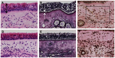
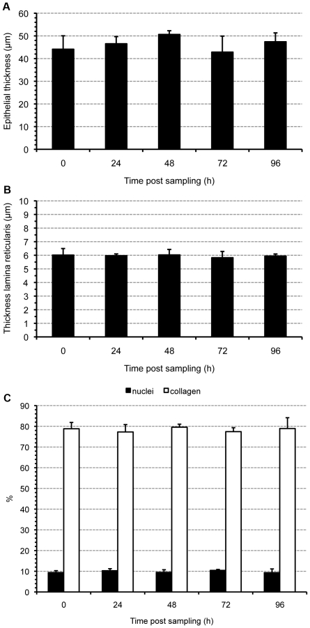
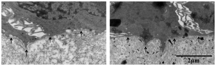
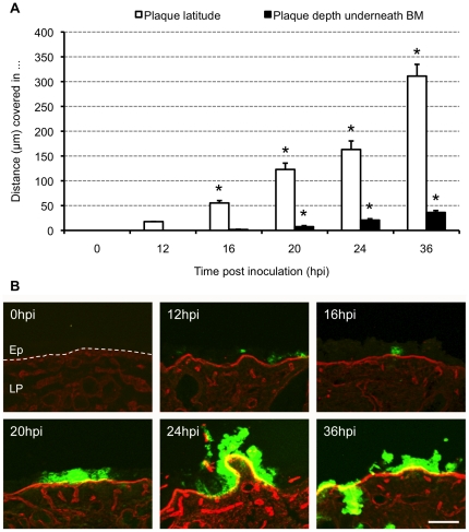
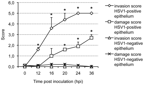
Methodology/principal findings: A human model consisting of nasal respiratory mucosa explants was characterised. Viability and integrity were examined during 96 h of cultivation. HSV1-mucosa interactions were analysed. In particular, we investigated whether HSV1 is able to reach the stroma. Explant viability and integrity remained preserved. HSV1 induced rounding up and loosening of epithelial cells with very few apoptotic and necrotic cells observed. Following 16-24 h of infection, HSV1 penetrated the basement membrane and replicated in the underlying lamina propria.

Conclusions/significance: This human explant model can be used to study virus-mucosa interactions and viral mucosal invasion mechanisms. Using this model, our results provide a novel insight into the HSV1 stromal invasion mechanism and for the first time directly demonstrate that HSV1 can penetrate the basement membrane.


Conflict of interest statement

Competing Interests: Reginald Clayton is employed by a commercial company, Tibotec BVBA. There are no patents, products in development or marketed products to declare. This does not alter the authors' adherence to all the PLoS ONE policies on sharing data and materials.

Figures

Figure 1
Figure 1. Light photomicrographs of the human model.
Representative light photomicrographs of human nasal respiratory mucosa explants at 0 (A, B, C) and 96 h (D, E, F) of in vitro cultivation are illustrated. Light microscopical morphometric analysis was performed to evaluate the maintenance of structural integrity of the explants during in vitro cultivation. The three-dimensional organization of the explants was assessed by evaluating the morphometry of the epithelium, lamina reticularis and lamina propria. Eight-micron-sections were stained with haematoxylin-eosin (A and D) for evaluation of the epithelial thickness (indicated by black arrows). A reticulin staining (B and E) was performed to measure the thickness of the lamina reticularis (indicated by white arrows). A Van Gieson staining (C and F) was used to count the relative amounts of collagen and nuclei within a region of interest (roi indicated by a rectangle) of the lamina propria. By setting a threshold, different colors were assigned to collagen and nuclei, respectively, and the percentages of collagen and nuclei were determined within this roi. Bar, 50 µm.
Figure 2
Figure 2. Morphometric evaluation of the human model.
Maintenance of the structural integrity of the explants during in vitro cultivation was evaluated by assessing the three-dimensional organization of the explants. Epithelial thickness (A), thickness of the lamina reticularis (B) and percentages of nuclei and collagen within a region of interest of the lamina propria (C) were evaluated in explants at different time points of in vitro cultivation. Data are represented as means+SD (error bars) of triplicate independent experiments.
Figure 3
Figure 3. Transmission electron photomicrographs of the human model.
The continuity of the lamina densa was evaluated during in vitro cultivation to assess the structural integrity of this basement membrane layer. The photomicrographs show representative images of the intact lamina densa in human nasal respiratory explants at 0 (A) and 96 (B) h of in vitro cultivation. Arrows indicate the lamina densa.
Figure 4
Figure 4. Evolution of HSV1 mucosal spread.
(A) Kinetic evolution of HSV1 plaque formation. Explants were inoculated with 1 ml virus suspension containing 107 TCID50/ml HSV1 VR-733 and sampled at 0, 12, 16, 20, 24 and 36 h post inoculation (pi). Serial 20 µm cryosections were made and plaque latitude (white bars) and plaque depth underneath the basement membrane (BM), distance covered by HSV1 in the lamina propria, (black bars) were measured using ImageJ. Data are represented as means of 10 plaques of triplicate independent experiments+SD (error bars). *, Significant differences at the 0.05 level. (B) Representative confocal photomicrographs of the evolution of HSV1 VR-733 spread in human nasal respiratory explants at 0, 12, 16, 20, 24 and 36 h pi. Collagen IV is visualised by red fluorescence. Green fluorescence visualises HSV1 antigens. Bar, 100 µm. Abbreviations: Ep, epithelium; LP, lamina propria. The BM is marked with a dashed line.
Figure 5
Figure 5. HSV1 invasion score and epithelial damage score.
Explants were inoculated with 1 ml 107 TCID50/ml HSV1 VR-733 and sampled at different time points post inoculation (pi). Serial 20 µm cryosections were made and evaluated. HSV1 mucosal invasion in the depth was graded on a 6-point scale, as follows: 0 = epithelium not infected, 1 = columnar cell(s) infected, 2 = suprabasal cell(s) infected, 3 = basal cell(s) infected, 4 = basement membrane and HSV1 colocalisation, HSV1 does not cross the basement membrane, 5 = HSV1 penetrates the basement membrane into the lamina propria. Epithelial damage was graded on a 4-point scale, as follows: 0 = no damage, 1 = superficial damage, 2 = epithelial damage involving basal cells, basal epithelial cells partly detached, 3 = epithelium severely damaged, loose. Both scales were combined on the same axis. The scores represent the mean of the scores of 10 different regions of HSV1-negative (100 cells) and HSV1-positive (virus plaque) cells per person at 0, 12, 16, 20, 24 and 36 h post inoculation (pi); experiments were performed in triplicate. Error bars indicate SD. *, Significant differences compared with the control (HSV1-negative epithelium) at the 0.05 level.

References

    1. Marshall DS, Linfert DR, Draghi A, McCarter YS, Tsongalis GJ. Identification of herpes simplex virus genital infection: comparison of a multiplex PCR assay and traditional viral isolation techniques. Mod Pathol. 2001;14:152–156. - PubMed
    1. Peña KC, Adelson ME, Mordechai E, Blaho JA. Genital herpes simplex virus type 1 in women: detection in cervicovaginal specimens from gynecological practices in the United States. J Clin Microbiol. 2010;48:150–153. - PMC - PubMed
    1. Whitley RJ. Herpes simplex viruses. In: Knipe DM, Howley PM, Griffin DE, Lamb RA, Martint MA, et al., editors. Fields virology. Philadelphia: Lippincott, Williams, Wilkins; 2001. pp. 2461–2509.
    1. Fleming DT, McQuillan GM, Johnson RE, Nahmias AJ, Aral SO, et al. Herpes simplex virus type 2 in the United States, 1976 to 1994. N Engl J Med. 1997;337:1105–111. - PubMed
    1. Mitchell BM, Bloom DC, Cohrs RJ, Gilden DH, Kennedy PG. Herpes simplex virus-1 and varicella-zoster virus latency in ganglia. J Neurovirol. 2003;9:194–204. - PubMed

Publication types

LinkOut - more resources