. 2011 Oct;401(6):1881-8.
doi: 10.1007/s00216-011-5257-z. Epub 2011 Jul 26.

Kinetic analysis of PI3K reactions with fluorescent PIP2 derivatives

Affiliations

Kinetic analysis of PI3K reactions with fluorescent PIP2 derivatives

Weigang Huang et al. Anal Bioanal Chem. 2011 Oct.

Abstract

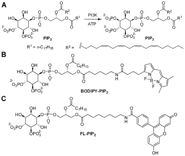
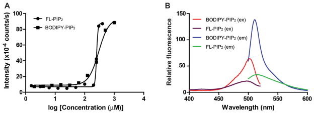
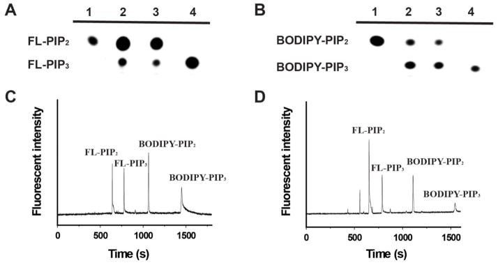
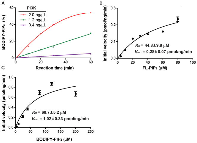
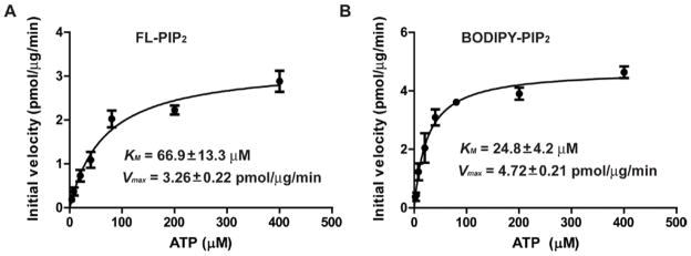
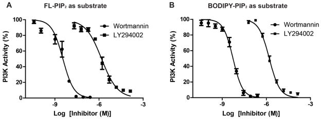
Phosphatidylinositol 3-kinase (PI3K) signaling plays important roles in cell differentiation, proliferation, and migration. Increased mutations and expression levels of PI3K are hallmarks for the development of certain cancers. Pharmacological targeting of PI3K activity has also been actively pursued as a novel cancer therapeutic. Consequently, measurement of PI3K activity in different cell types or patient samples holds the promise as being a novel diagnostic tool. However, the direct measurement of cellular PI3K activity has been a challenging task. We report here the characterization of two fluorescent PIP(2) derivatives as reporters for PI3K enzymatic activity. The reporters are efficiently separated from their corresponding PI3K enzymatic products through either thin layer chromatography (TLC) or capillary electrophoresis (CE), and can be detected with high sensitivity by fluorescence. The biophysical and kinetic properties of the two probes are measured, and their suitability to characterize PI3K inhibitors is explored. Both probes show similar capacity as PI3K substrates for inhibitor characterization, yet also possess distinct properties that may suggest their different applications. These characterizations have laid the groundwork to systematically measure cellular PI3K activity, and have the potential to generate molecular fingerprints for diagnostic and therapeutic applications.


Figures

Figure 1
Figure 1
Schematic illustration of the PI3K reaction. A. the endogenous PIP2 is phosphorylated at the 3′-OH to form PIP3; B, C. Chemical structures of fluorescent PIP2 derivatives BODIPY-PIP2 (B) and FL-PIP2 (C).
Figure 2
Figure 2
Biophysical characterization of FL-PIP2 and BODIPY-PIP2. A. Measurement of critical micelle concentrations (CMCs) of FL-PIP2 and BODIPY-PIP2. B. Fluorescence excitation and emission spectra of FL-PIP2 and BODIPY-PIP2.
Figure 3
Figure 3
Separation of the PI3K reaction mixtures with fluorescent PIP2 derivatives through TLC or CE analysis. A. the separation of the reaction mixture of FL-PIP2 on TLC. lane 1: FL-PIP2; lane 2: the reaction mixture was extracted with CHCl3/MeOH (v/v = 2:1) before TLC separation; lane 3: the reaction mixture was diluted with CHCl3/MeOH (v/v = 1:1) and separated on TLC; lane 4: FL-PIP3. B. the separation of the reaction mixture of BODIPY-PIP2 on TLC. lane 1: BODIPY-PIP2; lane 2: the reaction mixture was extracted with CHCl3/MeOH (v/v = 2:1) before TLC separation; lane 3: the reaction mixture was diluted with CHCl3/MeOH (v/v = 1:1) and separated on TLC; lane 4: BODIPY-PIP3. C. The separation of a mixture of FL-PIP2, FL-PIP3, BODIPY-PIP2, and BODIPY-PIP3 by CE; D. A 1:1 mixture of FL-PIP2 and BODIPY-PIP2 were used for the PI3K reaction, and the reaction mixture was separated by CE.
Figure 4
Figure 4
Kinetic analysis of PI3K reaction with fluorescent PIP2 derivatives. A. The reaction progress curves with different amounts of PI3K were used. B. Kinetic studies on PI3K reaction with FL-PIP2. C. Kinetic studies on PI3K reaction with BODIPY-PIP2. Each experiment was carried out in duplicate and repeated three times.
Figure 5
Figure 5
Measurement of KM for ATP for the PI3K reactions with FL-PIP2 (A) or BODIPY-PIP2 (B) as the lipid substrate. Each experiment was carried out in duplicate and repeated three times.
Figure 6
Figure 6
Dose response curves of PI3K inhibitors LY294002 and wortmannin. A. IC50 values of LY294002 and wortmannin were measured with FL-PIP2 as the PI3K substrate; B. IC50 values of LY294002 and wortmannin were measured with BODIPY-PIP2 as the PI3K substrate. Each experiment was carried out in duplicate and repeated three times.

Similar articles

Cited by

References

    1. Di Paolo G, De Camilli P. Phosphoinositides in cell regulation and membrane dynamics. Nature. 2006;443:651–657. - PubMed
    1. Balla T, Szentpetery Z, Kim YJ. Phosphoinositide signaling: new tools and insights. Physiology (Bethesda) 2009;24:231–244. - PMC - PubMed
    1. Castellino RC, Durden DL. Mechanisms of disease: the PI3K-Akt-PTEN signaling node--an intercept point for the control of angiogenesis in brain tumors. Nat Clin Pract Neurol. 2007;3:682–693. - PubMed
    1. Wymann MP, Schneiter R. Lipid signalling in disease. Nat Rev Mol Cell Biol. 2008;9:162–176. - PubMed
    1. Liu ZN, Roberts TM. Human tumor mutants in the p110 alpha subunit of PI3K. Cell Cycle. 2006;5:675–677. - PubMed

Publication types

MeSH terms

Substances

LinkOut - more resources