. 2011 Dec;35(12):2128-38.
doi: 10.1111/j.1530-0277.2011.01577.x. Epub 2011 Jul 25.

Elevated activation of ERK1 and ERK2 accompany enhanced liver injury following alcohol binge in chronically ethanol-fed rats

Affiliations

Elevated activation of ERK1 and ERK2 accompany enhanced liver injury following alcohol binge in chronically ethanol-fed rats

Annayya R Aroor et al. Alcohol Clin Exp Res. 2011 Dec.

Abstract

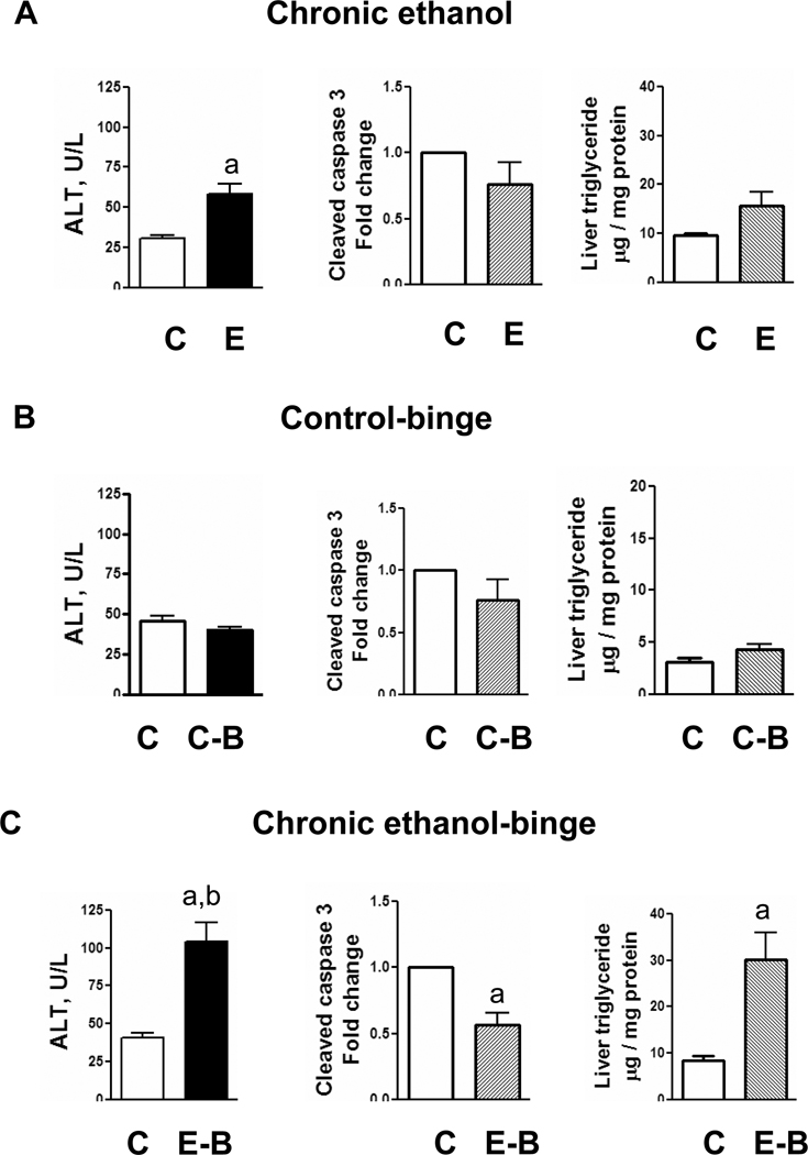
Background: Binge drinking after chronic ethanol consumption is one of the important factors contributing to the progression of steatosis to steatohepatitis. The molecular mechanisms of this effect remain poorly understood. We have therefore examined in rats the effect of single and repeat ethanol binge superimposed on chronic ethanol intake on liver injury, activation of mitogen-activated protein kinases (MAPKs), and gene expression.

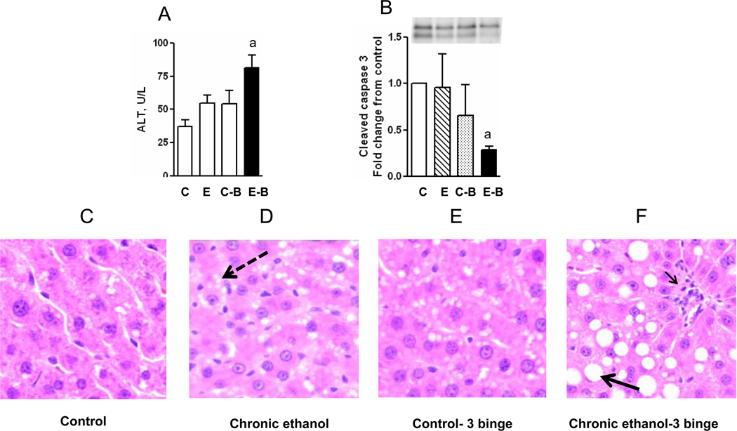
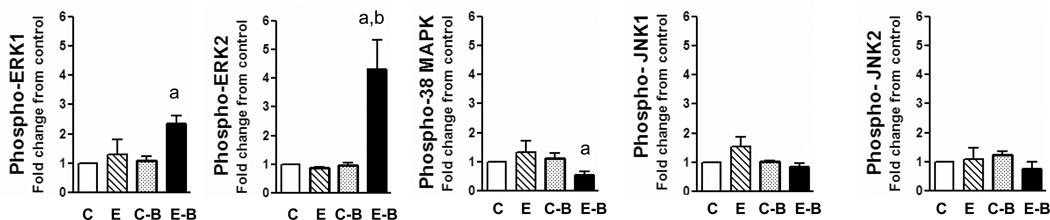
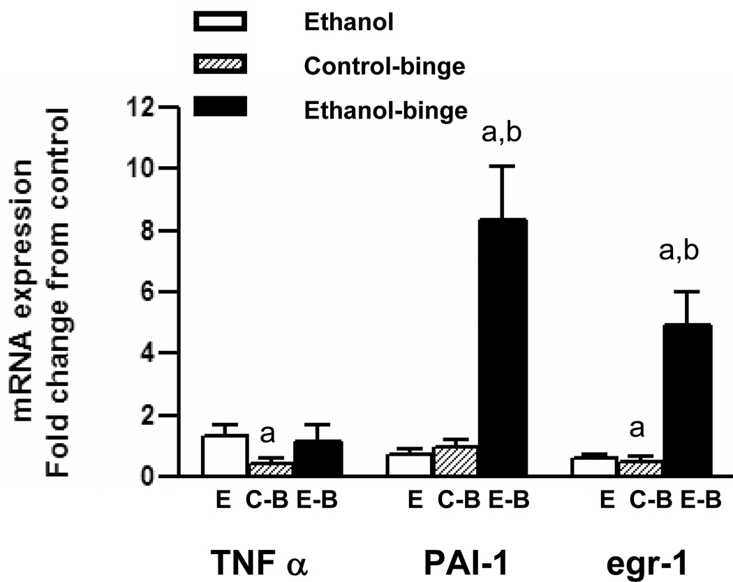
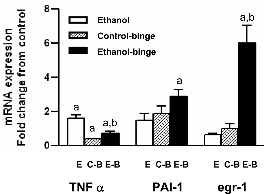
Methods: Rats were chronically treated with ethanol in liquid diet for 4 weeks followed by single ethanol binge (5 gm/kg body weight) or 3 similar repeated doses of ethanol. Serum alcohol and alanine amino transferase (ALT) levels were determined by enzymatic methods. Steatosis was assessed by histology and hepatic triglycerides. Activation of MAPK, 90S ribosomal kinase (RSK), and caspase 3 were evaluated by Western blot. Levels of mRNA for tumor necrosis factor alpha (TNFα), early growth response-1 (egr-1), and plasminogen activator inhibitor-1 (PAI-1) were measured by real-time qRT-PCR.

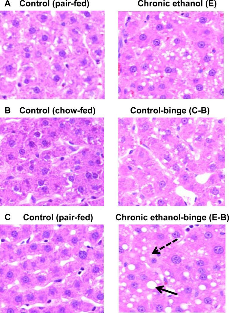
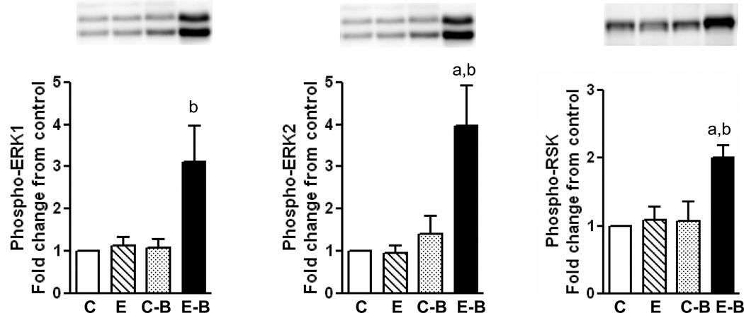
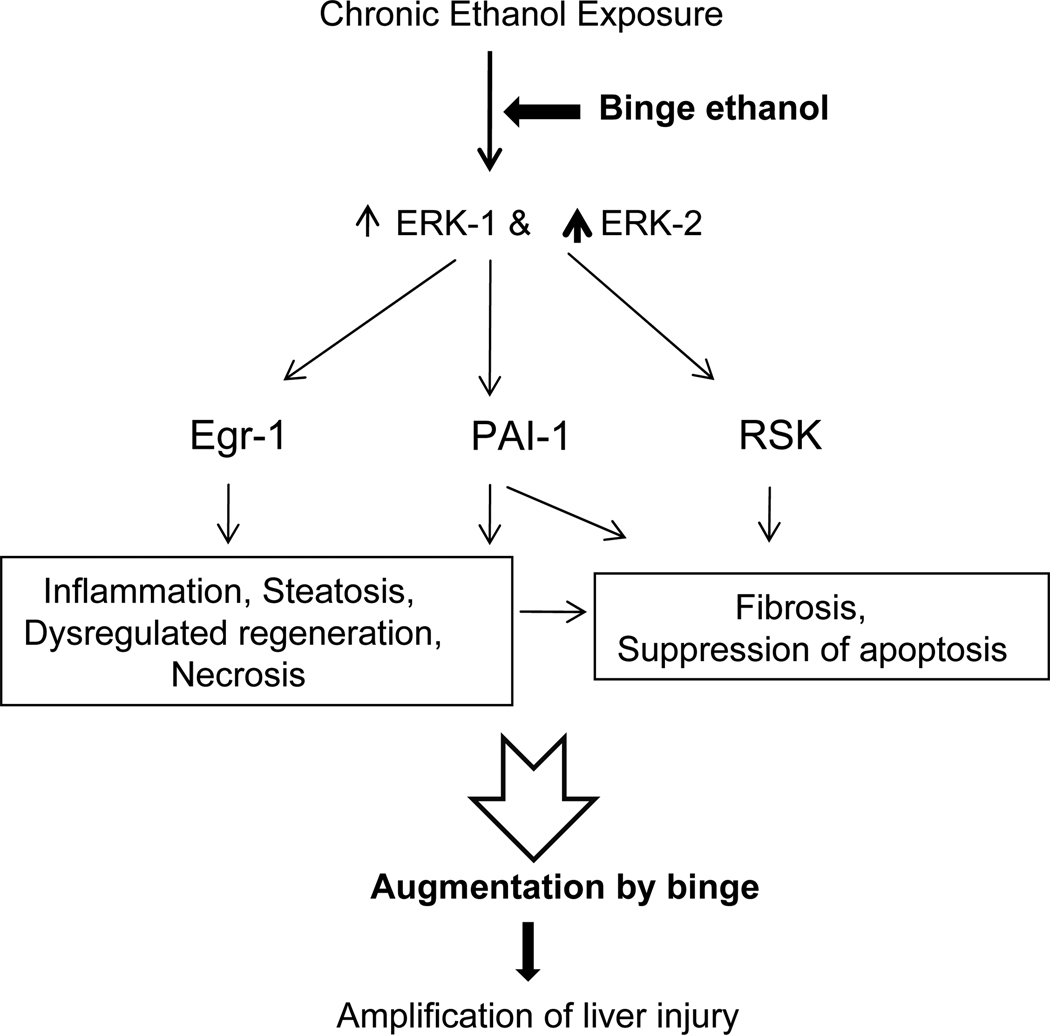
Results: Chronic ethanol treatment resulted in mild steatosis and necrosis, whereas chronic ethanol followed by binge group exhibited marked steatosis and significant increase in necrosis. Chronic binge group also showed significant increase (compared with chronic ethanol alone) in the phosphorylation of extracellular regulated kinase 1 (ERK1), ERK2, and RSK. Phosphorylation of c-Jun N-terminal kinase (JNK) and p38 MAPK did not increase by the binge. Ethanol binge, after chronic ethanol intake, caused increase in mRNA for egr-1 and PAI-1, but not TNFα.

Conclusions: Chronic ethanol exposure increases the susceptibility of rat liver to increased injury by 1 or 3 repeat binge. Among other alterations, the activated levels of ERK1, and more so ERK2, were remarkably amplified by binge suggesting a role of these isotypes in the binge amplification of the injury. In contrast, p38 MAPK and JNK1/2 activities were not amplified. These binge-induced changes were also reflected in the increases in the RNA levels for egr-1 and PAI-1. This study offers chronic followed by repeat binge as a model for the study of progression of liver injury by ethanol and highlights the involvement of ERK1 and ERK2 isotypes in the amplification of liver injury by binge ethanol.


Figures

Fig. 1
Fig. 1
Parameters of liver injury in chronic and chronic ethanol-single binge model. Rats were fed ethanol in liquid diet chronically for 4 weeks and then given a single ethanol binge dose (5 gm/kg). Samples were collected after 4 hrs. The levels of serum ALT, hepatic triglyceride and hepatic cleaved caspase-3 were determined as described under materials and methods. Values are mean ± SE (n=4 rats). Groups represent Chronic ethanol; Control-binge; Chronic ethanol-binge; a: significant from control group (p<0.05); b: significant from chronic ethanol group (p<0.05); C: Control; E: Chronic ethanol; C-B: Control-binge; E-B: Chronicethanol- binge. Control represents pair–fed animals for chronic and chronic-ethanol binge experiments. In control-binge experiments, the control represents chow-fed animals.
Fig. 2
Fig. 2
Histology of liver from chronic, and chronic ethanol-single binge treatment. Rats were fed ethanol in liquid diet chronically for 4 weeks and then given single ethanol binge dose (5 gm/kg). Samples were collected after 4 hrs. Sections of liver samples were stained with hematoxylin and eosin (x 200X). Values are mean ± SE (n=4 rats). Groups represent Chronic ethanol; Control-binge; C: Control; E: Chronic ethanol; C-B: Control-binge; E-B: Chronic-ethanol binge. Control represents pair–fed animals for chronic and chronic-ethanol binge experiments. In control-binge experiments, the control represents chow-fed animals. Solid arrow represents macrovesicular steatosis and broken arrow represents microvesicular steatosis.
Fig. 3
Fig. 3
Parameters of liver injury and histology in chronic and chronic ethanol- three binge model. Rats were fed ethanol in liquid diet chronically for 4 weeks and then given three binge (5 gm/kg) at 12 hr intervals. Four hr after the last dose, the levels of serum ALT, and hepatic cleaved caspase-3 were determined as described under materials and methods. Sections of liver samples were stained with hematoxylin and eosin. Control represents pair –fed animals and were given water for binge control. Values are mean ± SE (n=4 rats). Western blot represents a typical experiment. A: Serum ALT; B: hepatic cleaved caspase 3; C-F: Hemotoxylin and eosin staining (x 200X); a: significant from control group (p<0.05); C: Control (pair fed); E: Chronic ethanol; C-B: Control- ethanol binge; E-B: Chronic-ethanol binge. Large solid arrow represents macrovesicular steatosis. Large broken arrow represents microvesicular steatosis. Small solid arrow represents leukocyte infiltration.
Fig. 4
Fig. 4
Levels of phosphorylated ERK1/2, p38 MAPK and JNK1/2 in chronic and chronic-single ethanol binge treated rats. The chronic ethanol feeding (4 weeks) and binge (single) treatment was as in Fig. 1. The whole cell extracts from liver were subjected to western blotting with respective antibodies followed by densitometry of bands (see methods). Values are mean ± SE (n=4 rats). a: significant compared to control (p<0.05); b: significant from chronic ethanol group (p<0.05); C: Control; E: Chronic ethanol; C-B: Control- binge; E-B: Chronicethanol-binge. Control represents pair-fed animals for chronic and chronic-ethanol binge experiments. In control-binge experiments, the control represents chow-fed animals.
Fig. 5
Fig. 5
Levels of phosphorylated ERK1/2 and RSK in chronic and chronic-three ethanol binge treated rats. The chronic ethanol feeding (4 weeks) and three binge treatment was similar to Fig. 3. The whole cell extracts from liver were subjected to western blotting with respective antibodies followed by densitometry of bands (see methods). Values are mean ± SE (n=4 rats). A representative western blot is shown for each. a: significant compared to control (p<0.05); b: significant from chronic ethanol group (p<0.05); C: Control; E: Chronic ethanol; C-B: Control-binge; E-B: Chronic ethanol-binge.
Fig. 6
Fig. 6
TNFα, PAI-1 and egr-1 mRNA levels in chronic and chronic-ethanol single binge treated rats. After 4 weeks of chronic ethanol feeding, binge was administered as in Fig. 1. Total RNA was isolated from liver and reverse transcribed to cDNA. Aliquots of the cDNA preparations were amplified by real time qRT-PCR. The fold increase in mRNA levels was determined after normalizing the differences in level of GAPDH mRNA. Values are mean ± SE (n= 4 rats). a: significant compared to control (p<0.05); b: significant from chronic ethanol group (p<0.05); E: Chronic ethanol; C-B: Control- binge; E-B: Chronic –ethanol binge. Control represents pair–fed animals for chronic and chronic-ethanol binge experiments. In control-binge experiments, the control represents chow-fed animals.
Fig. 7
Fig. 7
TNFα, PAI-1 and egr-1 mRNA levels in chronic and chronic-ethanol three binge treated rats. After 4 weeks of chronic ethanol feeding, three binge was administered as in Fig. 3. Total RNA was isolated from liver and reverse transcribed to cDNA. Aliquots of the cDNA preparations were amplified by real time qRT-PCR. The fold increase in mRNA levels was determined after normalizing the differences in level of GAPDH mRNA. Values are mean ± SE (n= 4 rats). a: significant compared to control (p<0.05); b: significant from chronic ethanol group (p<0.05); E: Chronic ethanol; C-B: Control- ethanol binge; E-B: Chronic-ethanol binge.
Fig. 8
Fig. 8
A schematic diagram showing role of activation of ERK1, ERK2 isoforms, and RSK in the progression of liver injury by ethanol binge superimposed on chronic ethanol intake in the rat model. The scheme shows that chronic ethanol treatment sensitizes liver to binge ethanol induced enhancement of liver injury. The consequences of binge, after chronic ethanol intake, on dysregulated regeneration and fibrosis are speculative.

References

    1. Adachi M, Brenner DA. Clinical syndromes of alcoholic liver disease. Dig Dis. 2005;23:255–263. - PubMed
    1. Amersi F, Shen Xiu-Da, Anselmo D, Anselmo D, Melinek J, Iyer S, Southard DJ, Katori M, Volk HD, Busuttil RW, Buelow R, Kupiec-Weglinski JW. Ex vivo exposure to carbon monoxide prevents hepatic ischemia /reperfusion injury through p38 MAP kinase pathway. Hepatology. 2002;35:815–823. - PubMed
    1. Anjum R, Blenis J. The RSK family of kinases: emerging roles in cellular signalling. Nat Rev Mol Cell Biol. 2008;9:747–758. - PubMed
    1. Apte M, McCarroll J, Pirola R, Wilson J. Pancreatic MAP kinase pathways and acetaldehyde. Novartis Found Symp. 2007;285:200–211. - PubMed
    1. Aroor AR, Custer GW, Weng YI, Lee YJ, Shukla SD. Phosphatidylethanol mimics ethanol modulation of p42/44 mitogen-activated protein kinase signalling in hepatocytes. Alcohol Alcohol. 2002;37:534–539. - PubMed

Publication types

MeSH terms

Substances