. 2011 Nov;13(11):1668-82.
doi: 10.1111/j.1462-5822.2011.01646.x. Epub 2011 Aug 24.

Dissection of a type I interferon pathway in controlling bacterial intracellular infection in mice

Affiliations

Dissection of a type I interferon pathway in controlling bacterial intracellular infection in mice

Juliane Lippmann et al. Cell Microbiol. 2011 Nov.

Abstract

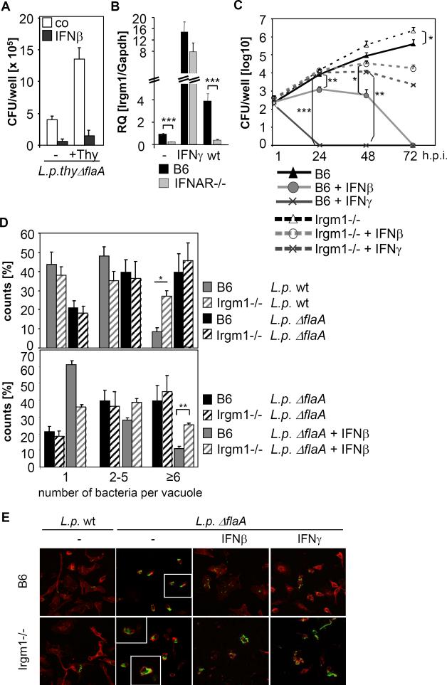
Defence mechanisms against intracellular bacterial pathogens are incompletely understood. Our study characterizes a type I IFN-dependent cell-autonomous defence pathway directed against Legionella pneumophila, an intracellular model organism and frequent cause of pneumonia. We show that macrophages infected with L. pneumophila produced IFNβ in a STING- and IRF3- dependent manner. Paracrine type I IFNs stimulated upregulation of IFN-stimulated genes and a cell-autonomous defence pathway acting on replicating and non-replicating Legionella within their specialized vacuole. Our infection experiments in mice lacking receptors for type I and/or II IFNs show that type I IFNs contribute to expression of IFN-stimulated genes and to bacterial clearance as well as resistance in L. pneumophila pneumonia in addition to type II IFN. Overall, our study shows that paracrine type I IFNs mediate defence against L. pneumophila, and demonstrates a protective role of type I IFNs in in vivo infections with intracellular bacteria.


Figures

Figure 1
Figure 1. Role of bacterial and host cell factors in stimulation of IFNβ expression in L. pneumophila-infected cells
(A) B6 BMMs were infected with L. pneumophila wt, mutants deficient for flaA, dotA and icmS or the thy mutant at a MOI of 25 for indicated time intervals. (B) B6 BMMs were treated with either bafilomycin A (Baf), chymostatin (Chy) or cytochalasin D (Cyt) or were left untreated and stimulated with viableor heat-killed (HK) L.p. wt for 4 hrs. (C) BMMs were transfected for 4 hrs with bacterial extracts that were either left untreated or were incubated with DNase I, RNase A, RNase H, DNase and RNases (D+R) or Proteinase K. (D, E) Wt BMMs and respective knockout BMMs were infected with L. pneumophila wt and/or ΔflaA (flaA) at a MOI of 25 for 4 hrs. (F-H) Wt or MAVS-/- BMMs were transfected with control siRNA (C) or siRNA against STING or DAI. After 48 hrs cells were infected with L. pneumophila for 4 hrs. (I) BMMs from wt and MAVS-/- mice were transfected with control siRNA (C) or siRNA against STING. After 48 hrs, cells were additionally transfected with Legionella DNA. Relative expression of IFNβ mRNA (A-E, H, I), STING (F) and DAI (G) was determined by quantitative RT-PCR. IFNβ mRNA values of untreated cells were set as 1. Data shown are representatives of at least three independent experiments carried out in triplicates. Unless indicated, statistics refer to the wt at the respective time point (A), to the untreated control (B) or to the untreated, transfected control (C) (*p<0.05; **p<0.01; ***p<0.001; relative to # no bacteria detected).
Figure 2
Figure 2. Type I IFNs activate a cell-autonomous defense independently of NAIP5/NLRC4-mediated recognition of flagellin and of IFNγ
(A-C) Intracellular growth of L.p. wt or ΔflaA in BMMs from wt (B6) and IRF3-/- (A), and IFNAR-/- (B, C) mice was monitored over 72 hrs by determining CFU per well at the indicated time points. BMMs were infected with L. pneumophila at a MOI of 0.1. Where indicated, cells were treated with 5 or 50 U/ml IFNβ 18 hrs prior and during the course of infection or were left untreated. Data shown are representatives of at least three independent experiments carried out in triplicates. (*p<0.05, **p<0.01, ***p<0.001, no indication if not significant)
Figure 3
Figure 3. Type I IFN affects bacterial numbers after establishment of the specialized Legionella-containing vacuole
(A,B) BMMs from B6 wt or IFNAR-/- mice were infected with L.p. wt or ΔflaA for 4 and 10 hrs and treated with IFNβ where indicated. (A) The percentage of vacuoles containing 1, 2-5 or more than 6 bacteria was determined from three individually performed experiments carried out in duplicates (*p<0.05, **p<0.01, no indication if not significant). (B) Representative images of Z-stack projections of macrophages infected with L.p. ΔflaA for 10 hrs are depicted. Zoom demonstrates individual vacuoles containing 1, 2-5 or more than 6 bacteria that were counted and classified. Scale bars correspond to 10 μm. (C, D) LCVs were isolated from untreated or IFNβ-stimulated wt (B6) or IFNAR-/- BMMs that were infected with GFP-expressing L.p. wt, ΔflaA or ΔdotA. LCVs were stained with antibodies against Calnexin or Lamp-1, respectively, and visualized by fluorescence microscopy. Results shown in (C) comprise data from three (Calnexin) and two (Lamp-1) individually performed experiments. Scale bars correspond to 5 μm (D).
Figure 4
Figure 4. The type I IFN-dependent resistance pathway inhibits replicating and non-replicating L. pneumophila, and is partly dependent on Irgm1
(A) BMMs were treated with 100 U/ml IFNβ as indicated, infected with L.p. thy ΔflaA in the presence or absence of supplemented thymidin, and intracellular bacterial numbers were determined 10 hrs p.i.. Data shown are representative of three independent experiments carried out in triplicates. (B) B6 and IFNAR-/- BMMs were stimulated with IFNγ or infected with L.p. wt for 24 hrs. Relative Irgm1 mRNA levels were quantified by Q-PCR. (C) BMMs from wt and Irgm1-/- mice were treated with IFNβ or IFNγ prior and during infection as indicated, and infected with L.p. ΔflaA. Bacterial replication was monitored over 72 hrs by determining CFU per well at the indicated time points. Data shown are representatives of four independent experiments carried out in triplicates. (D) BMMs from B6 and Irgm1-/- mice were treated with IFNβ as indicated prior and during infection, infected with L.p. wt or ΔflaA for 10 hrs, and the percentage of vacuoles containing 1, 2-5 or more than 6 bacteria was determined. Results shown comprise data from at least three individually performed experiments carried out in duplicates. (E) BMMs from B6 and Irgm1-/- mice were treated with IFNβ or IFNγ as indicated, and infected with L.p. wt or ΔflaA for 10 hrs. Bacteria were stained with an anti-Legionella-LPS antibody (green) and actin with phalloidin (red), visualization was done by fluorescence microscopy with an 63x objective. Inlets depict zoom of individual vacuoles. (*p<0.05, **p<0.01, ***p<0.001, no indication if not significant)
Figure 5
Figure 5. Expression of IFNβ and ISGs in L. pneumophila lung infection
Wt, IFNAR-/-, IFNGR-/- and IFNAR/IFNGR-/- mice were intranasally infected with L.p. ΔflaA (1×10⁶ bacteria/mouse) or received PBS instillation respectively. Mice were sacrificed on day 2, 4 or 6 post infection or 2 days after PBS instillation, and IFNβ (A), Irgm1 (B), Irga6 (C) and ISG15 (D) mRNA expression levels in lungs of infected mice were determined by quantitative RT-PCR relative to PBS-treated mice that were set as 1 (not shown) (n=4). (*p<0.05, **p<0.01, ***p<0.001, no indication if not significant)
Figure 6
Figure 6. Protective role of type I IFNs in L. pneumophila lung infection
Wt, IFNAR-/-, IFNGR-/- and IFNAR/IFNGR-/- mice were intranasally infected with L.p. ΔflaA (1×10⁶ bacteria/mouse) or received PBS instillation, respectively. Mice were sacrificed on day 2, 4 or 6 post infection or 2 days after PBS instillation, and bacterial loads in the lung (A) and spleen (B) were quantified. Gain/Loss of bodyweight post infection was determined at day 6 (C). Neutrophils in the lung (D) and in the blood (E) were determined at day 2, 4 and 6 post infection and 2 days after PBS instillation. (n=4; *p<0,05; # no bacteria detected).
Figure 7
Figure 7
Overview of the type I IFN-mediated cell-autonomous resistance pathway that controls L. pneumophila infection together with NAIP5/NLRC4 in vitro and together with NAIP5/NLRC4 and the type II IFN IFNγ in vivo.

References

    1. Ablasser A, Bauernfeind F, Hartmann G, Latz E, Fitzgerald KA, Hornung V. RIG-I-dependent sensing of poly(dA:dT) through the induction of an RNA polymerase III-transcribed RNA intermediate. Nat Immunol. 2009;10:1065–1072. - PMC - PubMed
    1. Amer A, Franchi L, Kanneganti TD, Body-Malapel M, Ozoren N, Brady G, Meshinchi S, Jagirdar R, Gewirtz A, Akira S, Nunez G. Regulation of Legionella phagosome maturation and infection through flagellin and host Ipaf. J Biol Chem. 2006 17 11;281:35217–35223. - PubMed
    1. Archer KA, Alexopoulou L, Flavell RA, Roy CR. Multiple MyD88-dependent responses contribute to pulmonary clearance of Legionella pneumophila. Cell Microbiol. 2009;11:21–36. - PMC - PubMed
    1. Auerbuch V, Brockstedt DG, Meyer-Morse N, O'Riordan M, Portnoy DA. Mice lacking the type I interferon receptor are resistant to Listeria monocytogenes. J Exp Med. 2004 16 8;200:527–533. - PMC - PubMed
    1. Barbalat R, Lau L, Locksley RM, Barton GM. Toll-like receptor 2 on inflammatory monocytes induces type I interferon in response to viral but not bacterial ligands. Nat Immunol. 2009;10:1200–1207. - PMC - PubMed

Publication types

MeSH terms