. 2011 Jul 28;71(2):319-31.
doi: 10.1016/j.neuron.2011.05.038.

Structure and assembly mechanism for heteromeric kainate receptors

Affiliations

Structure and assembly mechanism for heteromeric kainate receptors

Janesh Kumar et al. Neuron. .

Abstract

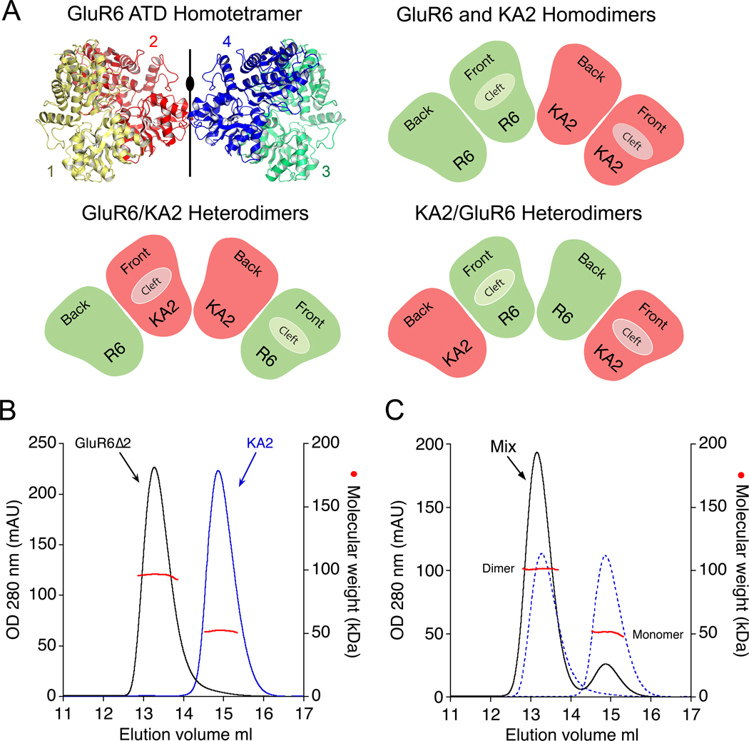
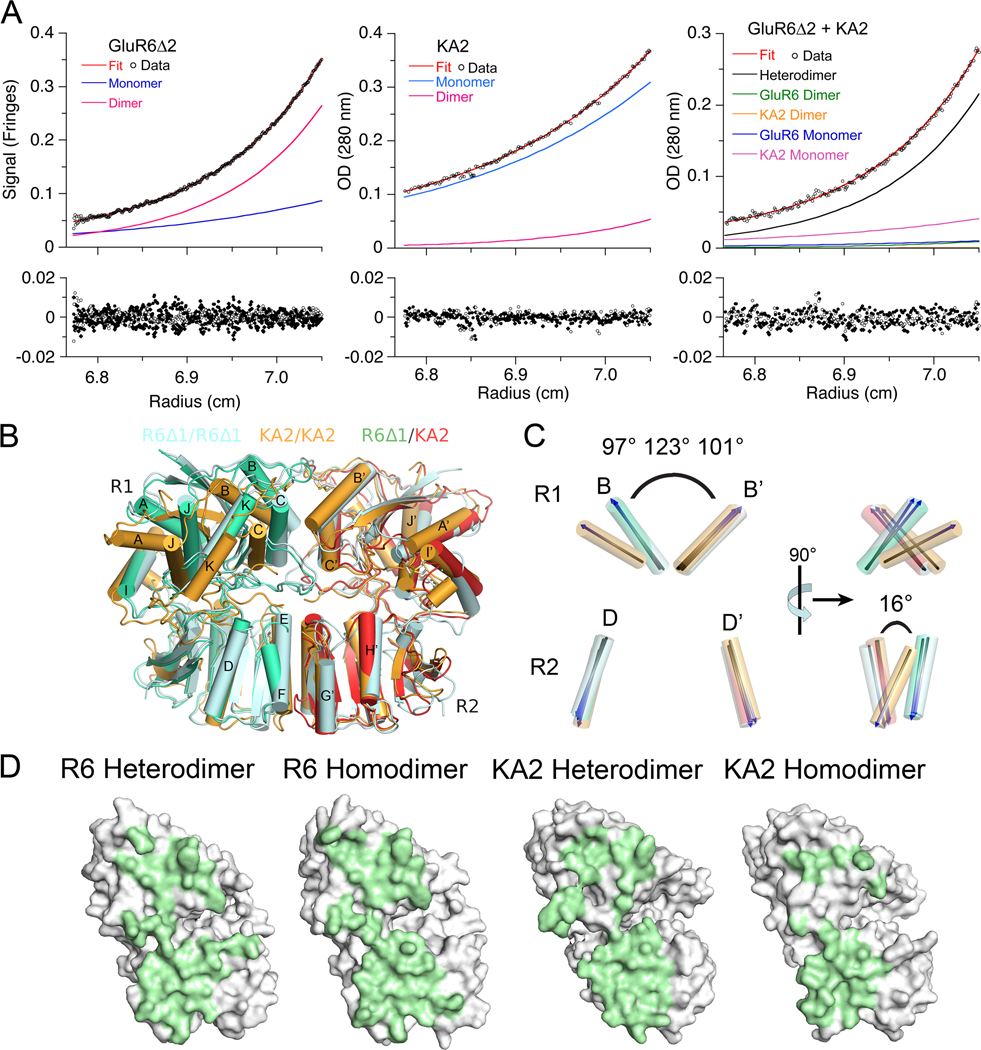
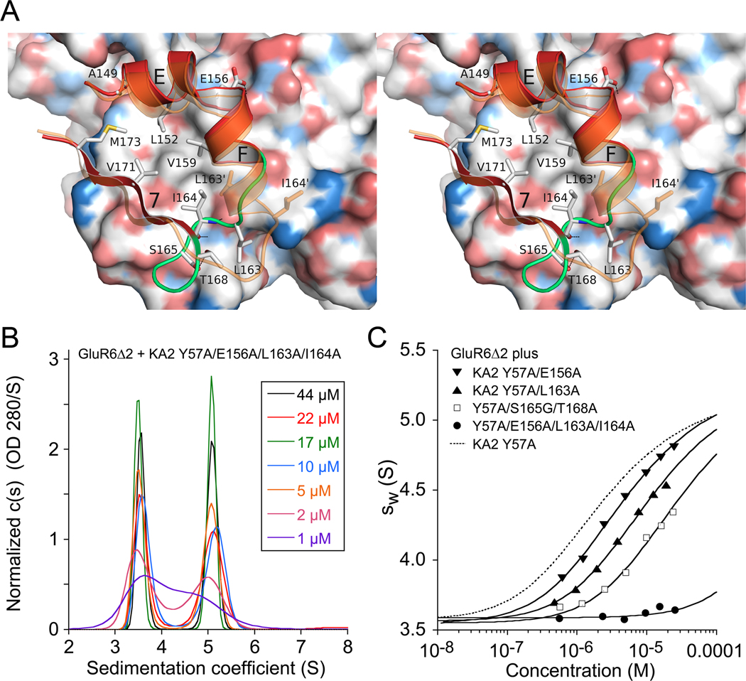
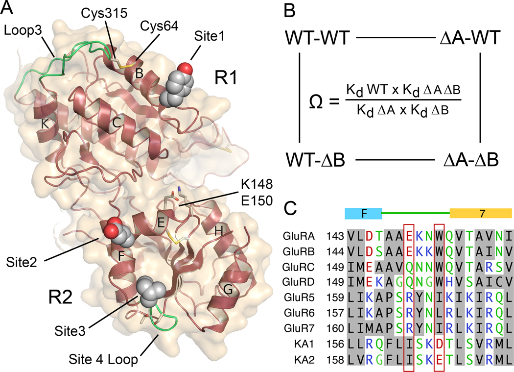
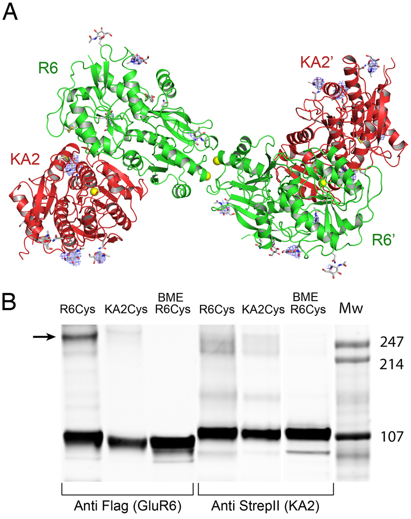
Native glutamate receptor ion channels are tetrameric assemblies containing two or more different subunits. NMDA receptors are obligate heteromers formed by coassembly of two or three divergent gene families. While some AMPA and kainate receptors can form functional homomeric ion channels, the KA1 and KA2 subunits are obligate heteromers which function only in combination with GluR5-7. The mechanisms controlling glutamate receptor assembly involve an initial step in which the amino terminal domains (ATD) assemble as dimers. Here, we establish by sedimentation velocity that the ATDs of GluR6 and KA2 coassemble as a heterodimer of K(d) 11 nM, 32,000-fold lower than the K(d) for homodimer formation by KA2; we solve crystal structures for the GluR6/KA2 ATD heterodimer and heterotetramer assemblies. Using these structures as a guide, we perform a mutant cycle analysis to probe the energetics of assembly and show that high-affinity ATD interactions are required for biosynthesis of functional heteromeric receptors.


Figures

Figure 1
Figure 1. Assembly principles for heteromeric glutamate receptor ATDs
(A) Crystal structure of the GluR6 ATD homotetramer (PDB 3H6H) with the four subunits individually colored, illustrating the global 2-fold axis of dimer symmetry; the cartoons show three possible subunit arrangements for an ATD heterotetramer assembled from GluR6 (green) and KA2 subunits (red). (B) Superimposed gel permeation chromatography profiles for the GluR6Δ2 and KA2 ATDs when the proteins were injected separately; analysis by SEC-UV/RI/MALS (red data points) revealed elution with mass values corresponding to dimers and monomers respectively. (C) When the two proteins were mixed at approximately equal concentrations prior to injection, the amplitude of the dimer peak increased, with a corresponding decrease in the monomer peak, indicating formation of GluR6/KA2 heterodimers; dashed lines show data from B scaled by 50% to account for the dilution factor when the samples were mixed.
Figure 2
Figure 2. The GluR6 and KA2 ATDs coassemble to form high affinity heterodimers
(A) Sedimentation equilibrium analysis for the GluR6Δ2 ATD, homodimer Kd 350 nM (left); the KA2 ATD, homodimer Kd 410 µM (middle); and an approximately equimolar mixture of the GluR6Δ2 and KA2 ATDs, heterodimer Kd 77 nM (right); fits to a monomer-dimer model (red line), with the calculated monomer and dimer populations shown as blue and pink lines, are shown for the three experiments (upper panels); the lower panels show residuals for single cells for a global fit to data for 3 loading concentrations each run at three speeds (6,500, 10,000 and 16,000 r.p.m). (B) The GluR6/KA2 heterodimer crystal structure superimposed using domain R2 coordinates on crystal structures for GluR6 (PDB 3G3H) and KA2 (PDB 3OM0) homodimers (C). Superimposed vectors drawn through helix B in domain 1 and helix D in domain 2, for the same structures, colored as in panel B, with the angle between helix B and its dimer partner indicated for the GluR6 homodimer, the KA2 homodimer, and the GluR6/KA2 heterodimer; rotation by 90° (right panel) reveals a 16° change in angle between helix D and helix D’ in domain R2 of the KA2 homodimer assembly. (D) Green shading illustrates buried molecular surfaces for the GluR6 and KA2 subunits for the same heterodimer and homodimer assemblies shown in B.
Figure 3
Figure 3. Role of conserved domain R1 aromatic side chains in heterodimer assembly
(A) Crystal structure of the GluR6Δ1/KA2 heterodimer domain R1 interface showing the interaction of KA2 Tyr57 with residues in α-helices B and C and loop three of the GluR6 subunit. A single subunit from the GluR6 homodimer crystal structure, superimposed on α-helices B and C, is colored light blue and shows the change in conformation of loop 3 and Asn 317 in the heterodimer assembly. (B) The view after rotation by 180° shows the interaction of GluR6 Phe58 with residues in α-helices B and C and loop three of the KA2 subunit. (C) Sedimentation velocity analysis for self assembly of the GluR6Δ2 F58A mutant ATD reveals a shift in monomer-dimer equilibrium due to a 2000-fold increase in Kd for homodimer formation; the profile for 36 µM GluR6Δ2 which sediments as a dimer is shown as a dashed line. (D) When the GluR6Δ2 F58A mutant is mixed with wt KA2 the sedimentation profile shifts to the right due to formation of ATD heterodimers; the profile for 14 µM KA2 which sediments as a monomer is shown as a dashed line. (E) A similar sedimentation profile was obtained for an equimolar mixture of the GluR6Δ2 and KA2 Y57A mutant ATDs. (F) Isotherms of weighted-average sedimentation coefficients fit with a monomer-dimer model reveals that compared to mutations in single subunits, mutation to alanine of both GluR6 F58 and KA2Y57 produces a 13-fold increase in Kd for heterodimer formation.
Figure 4
Figure 4. Domain R2 plays a key role in heterodimer assembly
(A) Stereo diagram of the GluR6/KA2 heterodimer domain R2 interface; the GluR6 subunit is shown as a molecular surface colored by atom type. The red ribbon diagram shows the KA2 subunit in the heterodimer assembly, with the loop connecting α-helix F with β-strand 7 shaded green; key residues forming intermolecular contacts are drawn as sticks; the ribbon diagram in transparent orange shows a subunit from a KA2 homodimer assembly. (B) Sedimentation velocity analysis for an equimolar mix of the GluR6Δ2 and KA2 E156A/L163A/I164A mutant ATDs reveals disruption of heterodimer assembly, with essentially no concentration dependence of the c(s) peak positions corresponding to monomers and dimers formed by the KA2 and GluR6 subunits respectively. (C) Isotherms of weighted-average sedimentation coefficients determined from peak integration of the c(s) data for individual KA2 Y57A domain R2 mutants mixed with GluR6Δ2 F58A; solid lines shows fits of a monomer-dimer model; the dashed line shows the isotherm for KA2 with wild type domain R2 residues.
Figure 5
Figure 5. Mutant cycle analysis for interactions between intermolecular contacts in the heterodimer assembly
(A) The KA2 subunit ATD viewed face on to the heterodimer surface, indicating the position of Tyr57 in site 1; Glu156 in site 2; Ile164 in site 3; Ser165 and Thr168 in site 4 that stabilizes the loop which makes site 3 contacts; the Cys64-Cys315 disulfide bond which holds loop 3 in place; and the location of Lys148 and Glu150. (B) Illustration of a mutant cycle for coupling between two sites indicated as A and B. (C) Amino acid alignment for AMPA and kainate receptors reveals a unique conservation of Ile164 in the KA2 subunits, and exchange of conserved residues in the loop connecting alpha helix F with beta strand 7.
Figure 6
Figure 6. Tetramer assembly is mediated by the GluR6 subunit
(A) Crystal structure of the GluR6/KA2 heterotetramer ATD assembly for wt GluR6 and KA2, colored green and red respectively; the ribbon diagram shows one of three identical tetramers for the 10 protomers in the asymmetric unit, with one tetramer formed by non crystallographic symmetry operations; side chains which support N-linked glycosylation are drawn as sticks; yellow spheres indicate the positions in the GluR6 and KA2 subunits at which cysteine mutations were introduced to test for formation of disulfide cross links in full length receptors. Electron density maps (Fo-Fc contoured at 3 σ blue mesh) illustrate features corresponding to glycan residues, which were not included in the model or used for refinement. (B) Western blots run under non-reducing conditions for detergent solubilized affinity-purified (StrepII tag) full-length heteromeric GluR6/KA2, in which Cys mutants were introduced at equivalent sites in the domain R2 lateral surface of either GluR6 or KA2 and probed against Flag (GluR6) and StrepII (KA2) epitopes; lanes 3 and 6 contain the same samples loaded in 1 and 4 but with the addition of 10 mM BME.
Figure 7
Figure 7. High affinity ATD interactions underlie assembly of heteromeric kainate receptors in vivo
(A) Responses to 60 µM glutamate, 500 µA AMPA, and 500 µM 5-iodowillardiine recorded at −60 mV from a Xenopus oocyte injected with 1.5 ng GluR6(R) and 3 ng wt KA2 cRNAs. (B) Responses for the same protocol but with the KA2 Y57A/E156A/L163A/I164A mutant. (C) Bar plot showing the mean ± sem for responses for wt KA2 (n=5) and the KA2 mutant (n=6).

Comment in

References

    1. Adams PD, Afonine PV, Bunkoczi G, Chen VB, Davis IW, Echols N, Headd JJ, Hung LW, Kapral GJ, Grosse-Kunstleve RW, et al. PHENIX: a comprehensive Python-based system for macromolecular structure solution. Acta Crystallogr D Biol Crystallogr. 2010;66:213–221. - PMC - PubMed
    1. Ayalon G, Segev E, Elgavish S, Stern-Bach Y. Two regions in the N-terminal domain of ionotropic glutamate receptor 3 form the subunit oligomerization interfaces that control subtype-specific receptor assembly. J Biol Chem. 2005;280:15053–15060. - PubMed
    1. Ayalon G, Stern-Bach Y. Functional assembly of AMPA and kainite receptors is mediated by several discrete protein-protein interactions. Neuron. 2001;31:103–113. - PubMed
    1. Brose N, Huntley GW, Stern-Bach Y, Sharma G, Morrison JH, Heinemann SF. Differential assembly of coexpressed glutamate receptor subunits in neurons of rat cerebral cortex. J Biol Chem. 1994;269:16780–16784. - PubMed
    1. Brown PH, Balbo A, Schuck P. Characterizing protein-protein interactions by sedimentation velocity analytical ultracentrifugation. Curr Protoc Immunol Chapter. 2008;18(Unit 18):15. - PubMed

Publication types

MeSH terms