. 2011 Oct;31(19):4052-62.
doi: 10.1128/MCB.05750-11. Epub 2011 Jul 26.

Balanced interactions between Lyn, the p85alpha regulatory subunit of class I(A) phosphatidylinositol-3-kinase, and SHIP are essential for mast cell growth and maturation

Affiliations

Balanced interactions between Lyn, the p85alpha regulatory subunit of class I(A) phosphatidylinositol-3-kinase, and SHIP are essential for mast cell growth and maturation

Peilin Ma et al. Mol Cell Biol. 2011 Oct.

Abstract

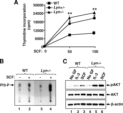
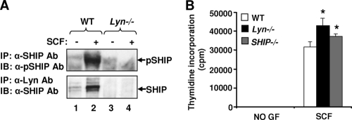
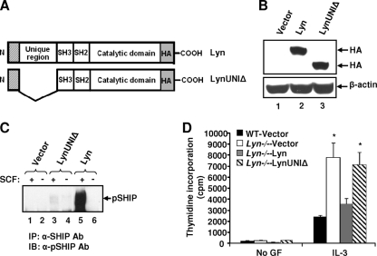
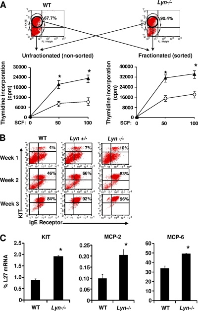
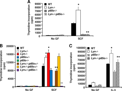
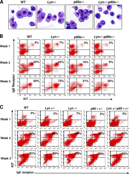
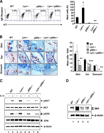
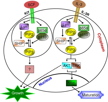
The growth and maturation of bone marrow-derived mast cells (BMMCs) from precursors are regulated by coordinated signals from multiple cytokine receptors, including KIT. While studies conducted using mutant forms of these receptors lacking the binding sites for Src family kinases (SFKs) and phosphatidylinositol-3-kinase (PI3K) suggest a role for these signaling molecules in regulating growth and survival, how complete loss of these molecules in early BMMC progenitors (MCps) impacts maturation and growth during all phases of mast cell development is not fully understood. We show that the Lyn SFK and the p85α subunit of class I(A) PI3K play opposing roles in regulating the growth and maturation of BMMCs in part by regulating the level of PI3K. Loss of Lyn in BMMCs results in elevated PI3K activity and hyperactivation of AKT, which accelerates the rate of BMMC maturation due in part to impaired binding and phosphorylation of SHIP via Lyn's unique domain. In the absence of Lyn's unique domain, BMMCs behave in a manner similar to that of Lyn- or SHIP-deficient BMMCs. Importantly, loss of p85α in Lyn-deficient BMMCs not only represses the hyperproliferation associated with the loss of Lyn but also represses their accelerated maturation. The accelerated maturation of BMMCs due to loss of Lyn is associated with increased expression of microphthalmia-associated transcription factor (Mitf), which is repressed in MCps deficient in the expression of both Lyn and p85α relative to controls. Our results demonstrate a crucial interplay of Lyn, SHIP, and p85α in regulating the normal growth and maturation of BMMCs, in part by regulating the activation of AKT and the expression of Mitf.


Figures

Fig. 1.
Fig. 1.
Deficiency of Lyn causes increased growth and enhanced PI3K activation in BMMCs. (A) BMMCs derived from WT, Lyn+/−, and Lyn−/− mice were starved in the absence of growth factors (GF) and subjected to a proliferation assay for 48 h. Shown is the amount of thymidine incorporation in an independent experiment performed in replicates of three with two different doses of SCF (50 and 100 ng/ml). The line graph represents the mean thymidine incorporation (counts per minute) ± the standard deviation in a representative experiment *, P < 0.05, Lyn+/− versus WT; **, P < 0.05, Lyn−/− versus WT. Similar results were observed in three independent experiments. (B and C) BMMCs from WT and Lyn−/− mice (5 × 10⁶) were starved, suspended in IMDM, and stimulated with cytokines, and lysates were analyzed for PI3K and AKT activation, respectively. Arrows indicate the levels of PI3P and pAKT in each lane. Similar results were observed in three independent experiments.
Fig. 2.
Fig. 2.
Lyn regulates the phosphorylation of SHIP in BMMCs. (A) Loss of SHIP activation due to Lyn deficiency in response to SCF stimulation in BMMCs. BMMCs derived from WT and Lyn−/− mice were starved and stimulated with SCF. Cell lysates were subjected to IP using an anti-SHIP or an anti-Lyn antibody (Ab). The immunoprecipitated (IP) protein complex was subjected to SDS-PAGE, and WB analysis was performed using an anti-phospho-SHIP (anti-pSHIP) or an anti-SHIP antibody. Arrows indicate the levels of phosphorylated and total SHIP, respectively, in each lane. Similar results were observed in two independent experiments. (B) Deficiency of SHIP in BMMCs mimics the enhanced growth observed in Lyn−/− cells. BMMCs derived from WT, Lyn−/−, and SHIP−/− mice were growth factor (GF) starved and subjected to a proliferation assay for 48 h. Shown is the amount of thymidine incorporation from an independent experiment performed in replicates of three in response to SCF. Bars represent the mean thymidine incorporation (counts per minute) ± the standard deviation in a representative experiment. *, P < 0.05, Lyn−/− and SHIP−/− versus WT. No significant difference in the growth of Lyn−/− versus SHIP−/− BMMCs was seen. Similar results were observed in three independent experiments.
Fig. 3.
Fig. 3.
The unique domain of Lyn regulates SHIP phosphorylation. (A) Schematic structures of the full-length WT version of Lyn and a mutant version of Lyn lacking its unique domain (LynUNIΔ). (B) Expression of WT Lyn and LynUNIΔ in Lyn-deficient BMMCs. Lyn−/− BMMCs were transduced with a retroviral construct expressing either the full-length WT version of Lyn or LynUNIΔ. EGFP-positive cells were sorted to homogeneity and used to perform WB analysis using an anti-HA antibody (upper panel) and β-actin antibody (lower panel). (C) Lyn−/− BMMCs expressing the empty vector (MIGR1), WT Lyn, or LynUNIΔ were starved of growth factors (GF) and stimulated with SCF. Lysates were subjected to IP using an anti-SHIP antibody (Ab), followed by WB analysis using an anti-phospho-SHIP (anti-pSHIP) antibody. Similar results were observed in two independent experiments. (D) Cells described in panels B and C were subjected to a proliferation assay for 48 h and pulsed with [3H]thymidine. Shown is the amount of thymidine incorporation in response to IL-3 from an independent experiment performed in replicates of three. Bars represent the mean thymidine incorporation (counts per minute) ± the standard deviation. *, P < 0.05, WT-Vector versus Lyn-Vector and LynUNIΔ. Similar results were obtained in four independent experiments.
Fig. 4.
Fig. 4.
Deficiency of Lyn in MCps results in enhanced BMMC maturation. (A) Enrichment of Lyn−/− KIT+ mast cell progenitors further enhances SCF-induced proliferation. Two- to 3-week-old MCps from WT and Lyn−/− BM were subjected to KIT staining and sorted to homogeneity for KIT expression. We subjected 5 × 10⁴ KIT+-sorted (fractionated) or nonsorted (unfractionated) MCps to SCF-induced proliferation for 48 h and pulsed them with [3H]thymidine. Shown is the amount of thymidine incorporation into unfractionated and fractionated [KIT-sorted] mast cell progenitors at two different concentrations of SCF (50 and 100 ng/ml). Lines denote the mean thymidine incorporation (counts per minute) ± the standard deviation from an independent experiment performed in replicates of three. *, P < 0.05, Lyn−/− versus WT. (B) BM cells from WT, Lyn+/−, and Lyn−/− mice were cultured under conditions that induce BMMC maturation. Cells were harvested weekly up to 3 weeks and stained with antibodies that recognize KIT and IgE receptor. Stained cells were analyzed by flow cytometry to determine the levels of KIT and IgE receptor expression at the end of each week. Shown are dot blot profiles of one of several experiments from the indicated genotypes analyzed at the end of weeks 1, 2, and 3. (C) Quantitative RT-PCR analysis of relative expression of mast cell markers associated with differentiation, including KIT, MCP-2, and MCP-6. Expression is normalized to L27. Values (percent L27 mRNA) are expressed as the mean results of experiments performed in triplicate. Error bars indicate the standard deviation. *, P < 0.001, WT versus Lyn−/−; n = 3.
Fig. 5.
Fig. 5.
Opposing roles for Lyn and p85α in BMMC growth. (A, B, and C) Loss of a single or two alleles of the p85α subunit of class IA PI3K in the setting of Lyn heterozygosity or homozygosity results in impaired BMMC growth. BMMCs from mice of the indicated genotypes were subjected to cytokine-induced proliferation for 48 h and pulsed with [3H]thymidine. Bars denote the average amounts of thymidine incorporation (counts per minute) ± the standard deviation in a representative experiment. Similar results were observed in three or four independent experiments. (A) *, P < 0.05, WT versus Lyn−/−; **, P < 0.05, Lyn−/− versus Lyn−/− p85α−/−. (B) *, P < 0.05, WT versus Lyn+/− and Lyn−/−; **, P < 0.05, Lyn+/− or Lyn−/− versus Lyn+/− p85α−/−, and Lyn+/− or Lyn−/− versus Lyn+/− p85α+/−. (C) *, P < 0.05, WT versus Lyn−/−; **, P < 0.05, Lyn−/− versus Lyn−/− p85α−/−. GF, growth factor.
Fig. 6.
Fig. 6.
Deficiency of p85α in Lyn−/− BM cells represses maturation of BMMCs. BMMCs were derived from WT, Lyn−/−, p85α−/−, and Lyn−/− p85α−/− mice. Cells were harvested after the indicated number of weeks and stained with Giemsa (A) or antibodies that recognize KIT and the IgE receptor (B). (A) Shown are representative cytospins of BMMC cultures derived from mice of the four genotypes. (B and C) Flow cytometric analysis demonstrating the expression of KIT and the IgE receptor in doubly positive cells at the indicated time points. The value in the upper right quadrant of each dot blot is the percentage of BM cells that are doubly positive for KIT and IgE receptor expression at the indicated times during culture. Similar findings were observed in BMMCs derived from two to four independent experiments.
Fig. 7.
Fig. 7.
Differential regulation of mast cells due to Lyn deficiency in vivo. (A) Cells from the peritoneal cavities of mice of the indicated genotypes were harvested and stained with antibodies against KIT and the IgE receptor. Representative dot blots indicate peritoneal cavity-derived mast cells from mice of the indicated genotypes stained with anti-KIT and anti-IgE receptor antibodies. The value in the upper right quadrant of each dot blot is the percentage of peritoneal cavity-derived cells that were doubly positive for KIT and IgE receptor expression. The bar graph represents a quantitative assessment of the total number of mast cells in 5 ml of peritoneal lavage fluid from mice of the indicated genotypes. Data are pooled from five independent experiments. Five to seven mice were used to generate the data in the left panel, and four to six mice were used to generate the data in the right panel. *, P < 0.05, WT versus Lyn−/−; **, P < 0.05, WT or Lyn−/− versus DKO. (B) Representative photomicrographs of toluidine blue-stained tissue sections derived from mice of the indicated genotypes. Arrows indicate tissue mast cells from mice of the indicated genotypes. The bar graph shows a quantitative assessment of toluidine blue-positive mast cells in the indicated tissues. n = 4 (mean ± standard deviation). *, P < 0.05, p85α−/− versus WT and Lyn−/−; **, P < 0.05, DKO versus WT and Lyn−/−; ***, P < 0.05, DKO versus WT. (C) Hyperactivation of AKT in Lyn−/− BMMCs is inhibited in the setting of p85α deficiency. BMMCs derived from mice of the indicated genotypes were starved in the absence of growth factors and stimulated with IL-3. Cell lysates were subjected to WB analysis using antibodies that recognize the activated versions of the indicated signaling proteins. The activation status of AKT and ERK1/2 is shown. Similar findings were obtained in two independent experiments. (D) Loss of Lyn expression in BMMCs results in enhanced expression of Mitf. BMMCs derived from WT, Lyn−/−, p85α−/−, and Lyn−/− p85α−/− mice were lysed and subjected to WB analysis using an anti-Mitf antibody. Arrows indicated the levels of expression of Mitf and β-actin (loading control) in each lane. Similar results were obtained in two independent experiments.
Fig. 8.
Fig. 8.
Schematic describing signaling via cytokine receptors in BMMCs involving SHIP, Lyn, and the p85α subunit of class IA PI3K. Upon ligand stimulation, the intracellular cytokine receptor domain is phosphorylated and recruits p85α to the receptor. This results in the generation of PIP3. PIP3 is hydrolyzed to PIP2 by SHIP, which regulates BMMC growth (in the case of KIT) and maturation (in the case of the IL-3 receptor). Our results show that the Lyn SFK regulates the activation of SHIP in MCps in response to cytokine stimulation by regulating its phosphorylation. Loss of Lyn expression in MCps results in a phenotype similar to that observed in MCps deficient in SHIP. The mechanism by which Lyn, SHIP, and p85α regulate mast cell maturation via the IL-3 receptor involves PI3K-mediated modulation of Mitf expression, which is associated with AKT and ERK1/2 activation. Furthermore, while loss of Lyn or SHIP in MCps results in similar functional outcomes via KIT and IL-3R, a requirement for p85α in KIT- and IL-3R-induced function(s) is distinct.

References

    1. Anderson S. M., Jorgensen B. 1995. Activation of src-related tyrosine kinases by IL-3. J. Immunol. 155:1660–1670 - PubMed
    1. Brown M. T., Cooper J. A. 1996. Regulation, substrates and functions of src. Biochim. Biophys. Acta 1287:121–149 - PubMed
    1. Costa J. J., Weller P. F., Galli S. J. 1997. The cells of the allergic response: mast cells, basophils, and eosinophils. JAMA 278:1815–1822 - PubMed
    1. Damen J. E., Ware M. D., Kalesnikoff J., Hughes M. R., Krystal G. 2001. SHIP's C-terminus is essential for its hydrolysis of PI3P and inhibition of mast cell degranulation. Blood 97:1343–1351 - PubMed
    1. Durand B., et al. 1994. Long-term generation of human mast cells in serum-free cultures of CD34+ cord blood cells stimulated with stem cell factor and interleukin-3. Blood 84:3667–3674 - PubMed

Publication types

MeSH terms

Substances

LinkOut - more resources