

MRI of tumor-associated macrophages with clinically applicable iron oxide nanoparticles
- PMID: 21791632
- PMCID: PMC3166957
- DOI: 10.1158/1078-0432.CCR-10-3420
MRI of tumor-associated macrophages with clinically applicable iron oxide nanoparticles
Abstract
Purpose: The presence of tumor-associated macrophages (TAM) in breast cancer correlates strongly with poor outcome. The purpose of this study was to develop a clinically applicable, noninvasive diagnostic assay for selective targeting and visualization of TAMs in breast cancer, based on magnetic resonanceI and clinically applicable iron oxide nanoparticles.
Experimental design: F4/80-negative mammary carcinoma cells and F4/80-positive TAMs were incubated with iron oxide nanoparticles and were compared with respect to magnetic resonance signal changes and iron uptake. MMTV-PyMT transgenic mice harboring mammary carcinomas underwent nanoparticle-enhanced magnetic resonance imaging (MRI) up to 1 hour and 24 hours after injection. The tumor enhancement on MRIs was correlated with the presence and location of TAMs and nanoparticles by confocal microscopy.
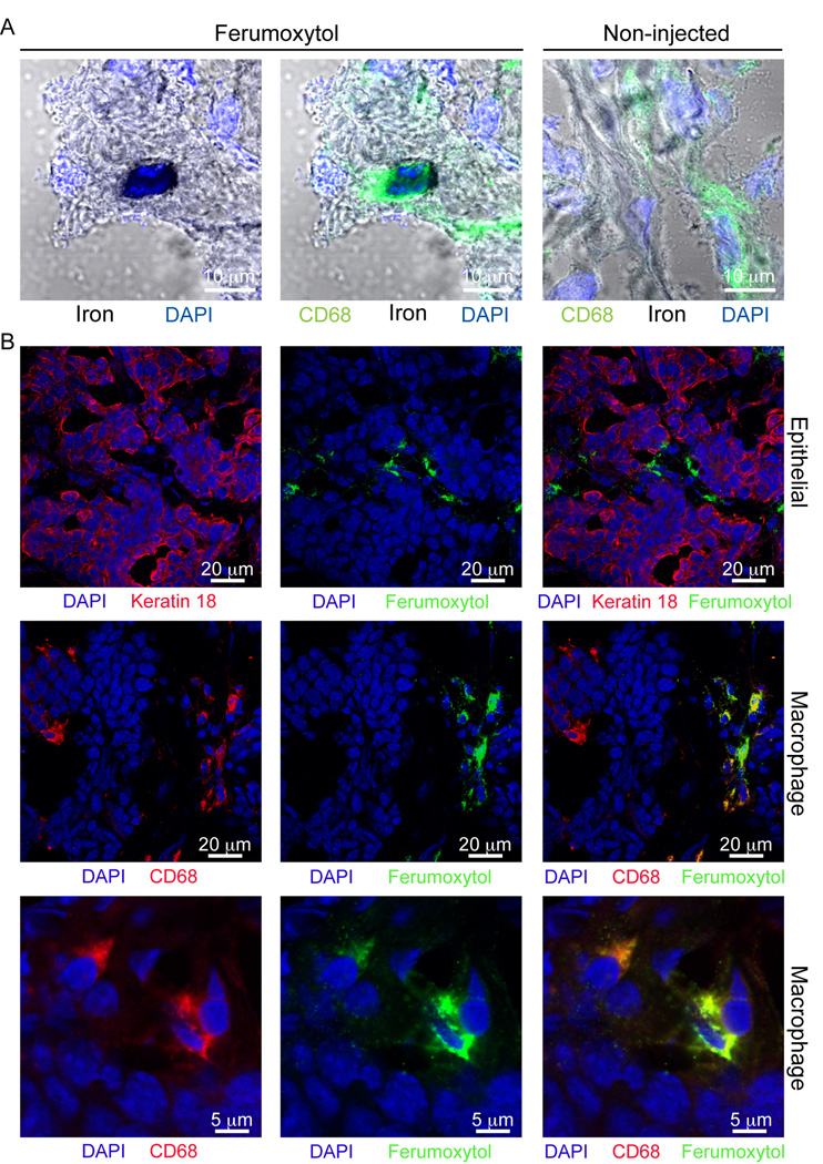
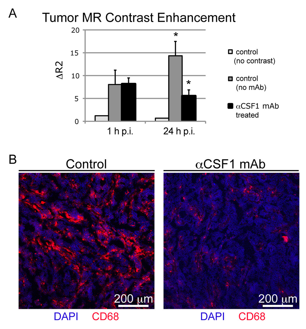
Results: In vitro studies revealed that iron oxide nanoparticles are preferentially phagocytosed by TAMs but not by malignant tumor cells. In vivo, all tumors showed an initial contrast agent perfusion on immediate postcontrast MRIs with gradual transendothelial leakage into the tumor interstitium. Twenty-four hours after injection, all tumors showed a persistent signal decline on MRIs. TAM depletion via αCSF1 monoclonal antibodies led to significant inhibition of tumor nanoparticle enhancement. Detection of iron using 3,3'-diaminobenzidine-enhanced Prussian Blue staining, combined with immunodetection of CD68, localized iron oxide nanoparticles to TAMs, showing that the signal effects on delayed MRIs were largely due to TAM-mediated uptake of contrast agent.
Conclusion: These data indicate that tumor enhancement with clinically applicable iron oxide nanoparticles may serve as a new biomarker for long-term prognosis, related treatment decisions, and the evaluation of new immune-targeted therapies.
©2011 AACR.
Figures


References
-
- de Visser KE, Eichten A, Coussens LM. Paradoxical roles of the immune system during cancer development. Nature Reviews Cancer. 2006;6:24–37. - PubMed
-
- Pollard JW. Tumour-educated macrophages promote tumour progression and metastasis. Nat Rev Cancer. 2004;4:71–78. - PubMed
-
- DeNardo DG, Johansson M, Coussens LM. Immune cells as mediators of solid tumor metastasis. Cancer Metastasis Rev. 2008;27:11–18. - PubMed
Publication types
MeSH terms
Substances
Grants and funding
- R01CA130980/CA/NCI NIH HHS/United States
- R01 CA132566/CA/NCI NIH HHS/United States
- P50 CA114747/CA/NCI NIH HHS/United States
- R21 CA156124/CA/NCI NIH HHS/United States
- U54 CA151459/CA/NCI NIH HHS/United States
- R01CA140943/CA/NCI NIH HHS/United States
- R01 CA130980/CA/NCI NIH HHS/United States
- R01 HD081123/HD/NICHD NIH HHS/United States
- P50 CA 58207/CA/NCI NIH HHS/United States
- R21CA156124/CA/NCI NIH HHS/United States
- R01CA132566/CA/NCI NIH HHS/United States
- R01 CA140943/CA/NCI NIH HHS/United States
- P50 CA58207/CA/NCI NIH HHS/United States
- P50 CA058207/CA/NCI NIH HHS/United States
LinkOut - more resources
Full Text Sources
Medical
