. 2011 Aug 15;204(4):592-600.
doi: 10.1093/infdis/jir352.

Critical role of serpinB1 in regulating inflammatory responses in pulmonary influenza infection

Affiliations

Critical role of serpinB1 in regulating inflammatory responses in pulmonary influenza infection

Dapeng Gong et al. J Infect Dis. .

Abstract

Background: Excessive inflammatory host response increases morbidity and mortality associated with seasonal respiratory influenza, and highly pathogenic virus strains are characterized by massive infiltration of monocytes and/or macrophages that produce a storm of injurious cytokines.

Methods: Here, we examined the role in respiratory influenza of serpinB1, an endogenous inhibitor of the serine proteases elastase, cathepsin G, and proteinase-3, increasingly recognized as regulators of inflammation.

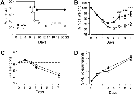
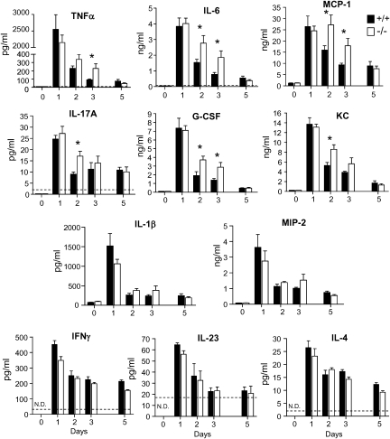
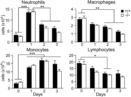
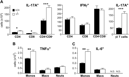
Results: After challenge with high-dose surfactant protein-D (SP-D)-sensitive influenza A/Philadelphia/82 (H3N2), serpinB1(-/-) mice died earlier and in greater numbers than did wild-type mice. Sublethally infected animals suffered increased morbidity, delayed resolution of epithelial injury, and increased immune cell death. Viral clearance and SP-D/SP-A upregulation were unimpaired and so were early virus-induced cytokine and chemokine burst and influx of large numbers of neutrophils and monocytes. Whereas initial cytokines and chemokines rapidly cleared in wild-type mice, TNF-α, IL-6, KC/CXCL1, G-CSF, IL-17A, and MCP-1/CCL2 remained elevated in serpinB1(-/-) mice. Monocyte-derived cells were the dominant immune cells in influenza-infected lungs, and those from serpinB1(-/-) mice produced excessive IL-6 and TNF-α when tested ex vivo. Pulmonary γδ T-cells that produced IL-17A were also increased.

Conclusions: Because viral clearance was unimpaired, the study highlights the critical role of serpinB1 in mitigating inflammation and restricting pro-inflammatory cytokine production in influenza infection.


Figures

Figure 1.
Figure 1.
Survival, morbidity, viral clearance and lung collectin levels of serpinB1−/− mice after influenza challenge. Wild type (+/+) and serpinB1 deficient (-/-) mice were inoculated with (A)107 ffc/mouse or (B-D) 2 ×10⁶ ffc/mouse of Phil/82. A, Kaplan-Meier survival curves for 12 each wild type and serpinB1−/− mice. B, Mean body weights were significantly different between genotypes by 2-way ANOVA with Bonferroni’s posttest (*, P < .05; ***, P < .001). Ten mice/genotype were evaluated of which 5 randomized mice/genotype were sacrificed after day 5. Data are representative of two experiments. C, Viral titers were determined by real time PCR in lung homogenates of mice sacrificed on the indicated days post-infection. The dotted line represents the inoculum. D, SP-D levels in lung homogenates were detected by western blot, quantified by densitometry and expressed as “μg equivalents” using recombinant rat SP-D as internal standard. Mean ± SEM for 4-5 mice/group, and the data were analyzed by 2-way ANOVA.
Figure 2.
Figure 2.
Lung histopathology, MPO activity, and death of recruited cells of influenza-infected mice. Wild type (+/+) and serpinB1 deficient (-/-) mice were infected with Phil/82 (2 × 10⁶ ffc/mouse) and were sacrificed on the indicated days. A, Hematoxylin and eosin-stained lung sections on day 2 and 3. Note that inflammatory cells with segmented and doughnut shaped nuclei characteristic of neutrophils and monocytes (arrowheads) are abundant in airways and peribronchiolar areas on day 2 in mice of both genotypes, together with substantial cellular debris in airways (single arrows). Monocytic cells with bean-shaped or round nuclei with a large cytoplasm (double arrows) suggestive of resolution were observed in airways and alveolar spaces of wild type mice on day 3 but were not observed in serpinB1−/− mice, where the histological picture remains with similar features as on day 2. Areas of the top panels (40x objective) were enlarged and are shown in the lower panels. Histology is representative of at least 3 mice/genotype/time point. B, MPO activity in lung homogenates. Mean ± SEM is shown for 4-5 mice/group, and the data were analyzed by 2-way ANOVA. C, ViDbright (dead) leukocytes (CD45+) in BAL. Mean ± SEM is shown for 4-5 mice/group, and data were analyzed by unpaired t test (*, P < .05).
Figure 3.
Figure 3.
Cytokines and chemokines in lungs of influenza-infected mice. Wild type (+/+) and serpinB1−/− mice were inoculated with sublethal dose Phil/82 and were sacrificed on the indicated days. Lung homogenates were assayed by ELISA for the indicated cytokine/chemokine. Mean ± SEM is shown for 3-5 mice/group, and data were analyzed by unpaired t test (*, P < .05). N.D., not detectable
Figure 4.
Figure 4.
Leukocyte subsets in lungs of influenza-infected mice. Wild type and serpinB1−/− mice were infected with sublethal dose Phil/82 and sacrificed on the indicated days. Subsets of leukocytes (CD45+) were quantified after enzymatic digestion of excised lungs. Shown are neutrophils (CD11b+Ly6G+), monocytes (CD11b+Ly6Gneg), macrophages (CD11bnegCD11c+ highly autofluorescent cells) and lymphocytes (low side scatter, CD11bneg). The number of cells for wild type and serpinB1−/− mice at each time point were analyzed by the unpaired t test; there were no differences between the genotypes. Changes of cell numbers over time were analyzed by one-way ANOVA followed by Tukey post test (*, P < .05; **, P < .01; ***, P < .0001). Mean ± SEM is shown for 4 mice/genotype/time point. Data are representative of 2–3 experiments.
Figure 5.
Figure 5.
IL-17A, IFN-γ, TNF-α, and IL-6 synthesizing lung leukocytes of infected mice. Wild type and serpinB1−/− mice were sacrificed on day 2 of sublethal Phil/82 infection. A, Single cell suspensions from excised lungs were cultured with PMA, ionomycin and brefeldin A and stained with the indicated antibodies and intracellularly for IL-17A and IFN-γ. IL-17A and IFN-γ producing cells were gated first on lymphocytes (low side scatter, CD45+CD11bneg). B, C, Single cell suspensions were incubated with LPS and brefeldin A and stained as in Figure 4 for monocytes, macrophages and neutrophils and intracellularly for (B) TNF-α or (C) IL-6. Mean ± SEM is shown for 4 mice/group, and data were analyzed by the unpaired t test (*, P < .05; **, P < .01; ***, P < .001; N.D., not detectable).

References

    1. Pham CT. Neutrophil serine proteases: specific regulators of inflammation. Nat Rev Immunol. 2006;6:541–50. - PubMed
    1. Cooley J, Takayama TK, Shapiro SD, Schechter NM, Remold-O'Donnell E. The serpin MNEI inhibits elastase-like and chymotrypsin-like serine proteases through efficient reactions at two active sites. Biochemistry. 2001;40:15762–70. - PubMed
    1. Benarafa C, Priebe GP, Remold-O'Donnell E. The neutrophil serine protease inhibitor serpinb1 preserves lung defense functions in Pseudomonas aeruginosa infection. J Exp Med. 2007;204:1901–9. - PMC - PubMed
    1. McGill J, Heusel JW, Legge KL. Innate immune control and regulation of influenza virus infections. J Leukoc Biol. 2009;86:803–12. - PMC - PubMed
    1. Tecle T, White MR, Gantz D, Crouch EC, Hartshorn KL. Human neutrophil defensins increase neutrophil uptake of influenza A virus and bacteria and modify virus-induced respiratory burst responses. J Immunol. 2007;178:8046–52. - PubMed

Publication types