

Macular features from spectral-domain optical coherence tomography as an adjunct to indirect ophthalmoscopy in retinopathy of prematurity
- PMID: 21792089
- PMCID: PMC3165115
- DOI: 10.1097/IAE.0b013e31821dfa6d
Macular features from spectral-domain optical coherence tomography as an adjunct to indirect ophthalmoscopy in retinopathy of prematurity
Abstract
Purpose: To compare vitreoretinal pathology imaged with portable handheld spectral-domain optical coherence tomography (SD-OCT) to conventional indirect ophthalmoscopic examination in neonates undergoing screening for retinopathy of prematurity.
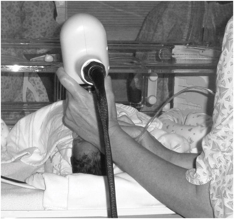
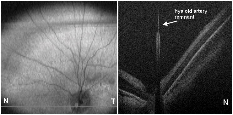
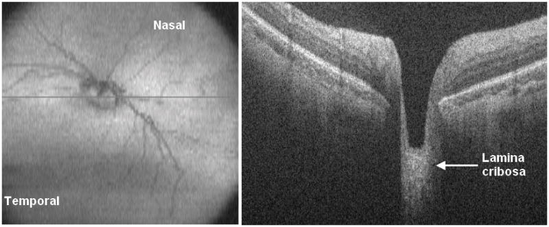
Methods: Spectral-domain optical coherence tomography images were collected from 76 eyes of 38 neonates during 118 routine retinopathy of prematurity examinations. Imaging sessions in the Neonatal Intensive Care Unit were performed immediately after the subjects underwent a standard ophthalmic examination with indirect ophthalmoscopic by a pediatric ophthalmologist. Masked certified SD-OCT graders evaluated scans for preretinal and retinal findings including material in the vitreous, epiretinal membrane, intraretinal cystoid structures and deposits, optic nerve and vascular features, and severity and location of retinopathy of prematurity. The frequency of detection of these features by clinical examination and evaluation of SD-OCT images was compared to determine potential clinical advantages for each modality.
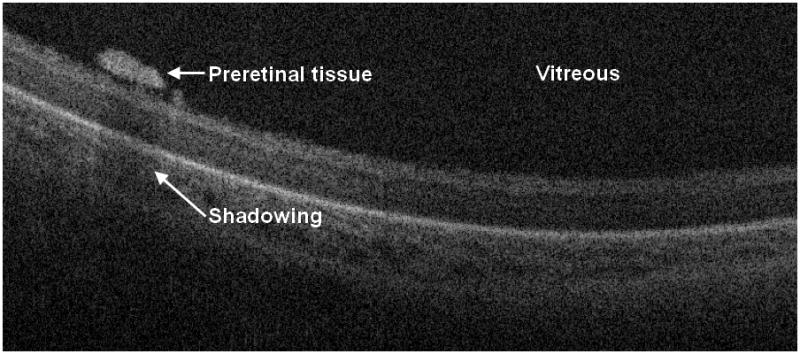
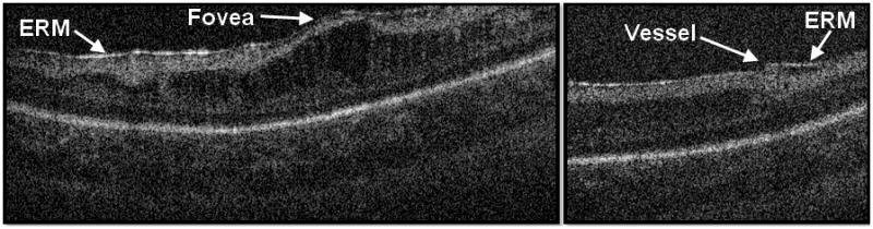
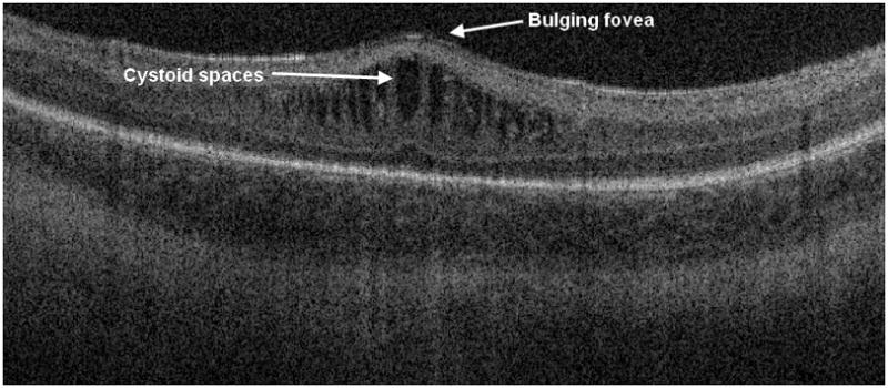
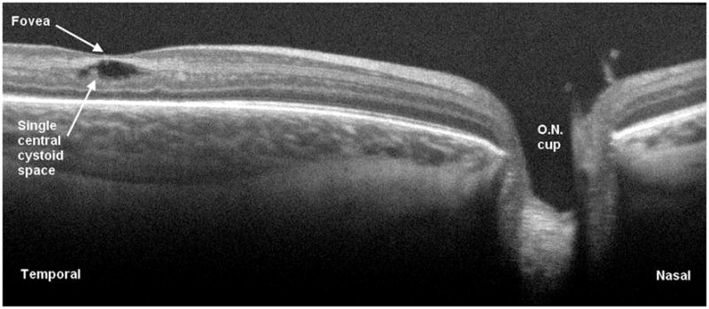
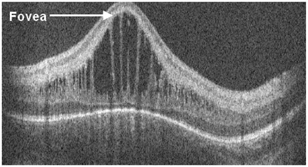
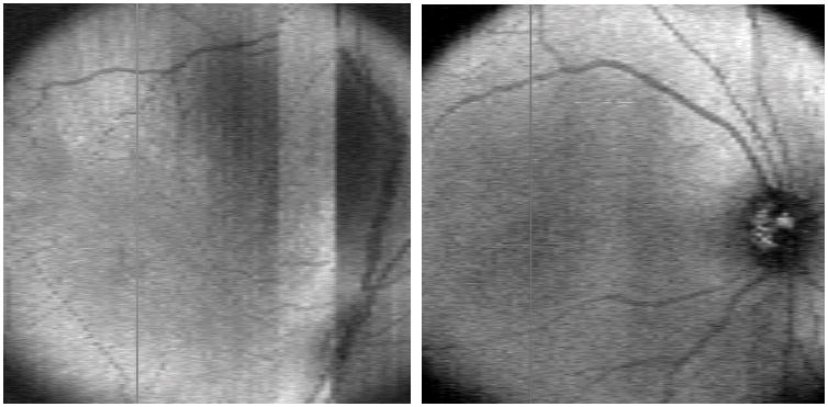
Results: Portable SD-OCT imaging characterized macular features of retinal cystoid structures in 39% of examinations and epiretinal membrane in 32% of examinations. Neither feature was visualized by indirect ophthalmoscopy in any cases. The clinician using indirect ophthalmoscopy detected stage of retinopathy of prematurity and the presence or absence of Plus or pre-Plus disease. These were not visualized with SD-OCT.
Conclusion: Spectral-domain optical coherence tomography provides new information about the premature infant retina that is of unknown importance relative to visual development and acuity. As used in this study, SD-OCT does not replace indirect ophthalmoscopy for evaluation of retinopathy of prematurity.
Figures
References
-
- Tasman W. Multicenter trial of cryotherapy for retinopathy of prematurity. Arch Ophthalmol. 1988;106(4):463–4. - PubMed
-
- Early Treatment For Retinopathy Of Prematurity Cooperative, G. Revised indications for the treatment of retinopathy of prematurity: results of the early treatment for retinopathy of prematurity randomized trial. Arch Ophthalmol. 2003;121(12):1684–94. - PubMed
-
- Screening examination of premature infants for retinopathy of prematurity. Pediatrics. 2001;108(3):809–11. - PubMed
-
- Mirza RG, Johnson MW, Jampol LM. Optical coherence tomography use in evaluation of the vitreoretinal interface: a review. Surv Ophthalmol. 2007;52(4):397–421. - PubMed
Publication types
MeSH terms
Grants and funding
LinkOut - more resources
Full Text Sources
Other Literature Sources
Research Materials
