. 2012 Jan;20(1):28-36.
doi: 10.1038/mt.2011.155. Epub 2011 Jul 26.

ACE2 and Ang-(1-7) confer protection against development of diabetic retinopathy

Affiliations

ACE2 and Ang-(1-7) confer protection against development of diabetic retinopathy

Amrisha Verma et al. Mol Ther. 2012 Jan.

Abstract

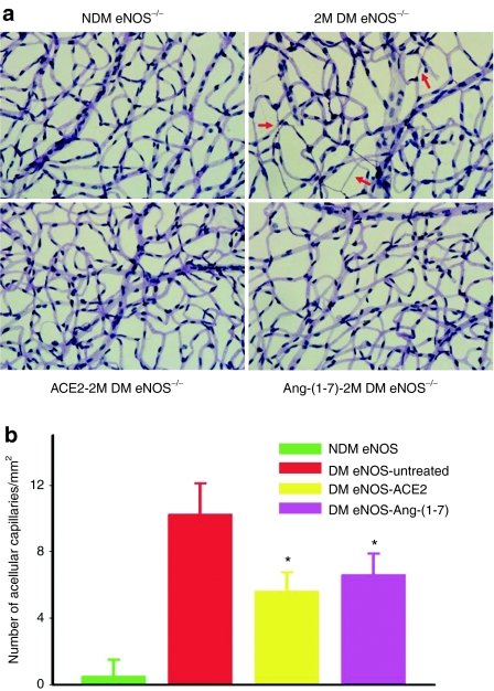
Despite evidence that hyperactivity of the vasodeleterious axis (ACE/angiotensin II (Ang II)/AT1 receptor) of the renin-angiotensin system (RAS) is associated with the pathogenesis of diabetic retinopathy (DR) use of the inhibitors of this axis has met with limited success in the control of this pathophysiology. We investigated the hypothesis that enhancing the local activity of the recently established protective axis of the RAS, ACE2/Ang-(1-7), using adeno-associated virus (AAV)-mediated gene delivery of ACE2 or Ang-(1-7) would confer protection against diabetes-induced retinopathy. Genes expressing ACE2 and Ang-(1-7) were cloned in AAV vector. The effects of ocular AAV-ACE2/Ang-(1-7) gene transfer on DR in diabetic eNOS(-/-) mice and Sprague-Dawley (SD) rats were examined. Diabetes was associated with approximately tenfold and greater than threefold increases in the ratios of ACE/ACE2 and AT1R/Mas mRNA levels in the retina respectively. Intraocular administration of AAV-ACE2/Ang-(1-7) resulted in significant reduction in diabetes-induced retinal vascular leakage, acellular capillaries, infiltrating inflammatory cells and oxidative damage in both diabetic mice and rats. Our results demonstrate that DR is associated with impaired balance of retinal RAS. Increased expression of ACE2/Ang-(1-7) overcomes this imbalance and confers protection against DR. Thus, strategies enhancing the protective ACE2/Ang-(1-7) axis of RAS in the eye could serve as a novel therapeutic target for DR.


Figures

Figure 1
Figure 1
Real-time reverse transcriptase (RT)-PCR analysis of retinal mRNA for renin-angiotensin system genes. Values on y-axis represent fold difference compared to age-matched nondiabetic retinal samples for each gene at each time point (14 day and 1 month after induced diabetes). DM, diabetic; NDM, nondiabetic. At least four eyes were analyzed at each time point. *P < 0.01 (versus NDM group).
Figure 2
Figure 2
Construction and characterization of adeno-associated virus (AAV) vectors expressing angiotensin-converting enzyme 2 (ACE2) and Ang-(1-7). (a) Maps of the AAV vector expressing the human ACE2 gene (hACE2) and the AAV vector expressing Ang-(1-7) gene. The Ang-(1-7) peptide is expressed as part of fusion protein, and cleaved upon secretion at the furin cleavage (FC) site. CBA, CMV-chicken-β-actin promoter; ITR, inverted terminal repeat. A control vector contains coding region for the secreted green fluorescent protein (GFP) without Ang-(1-7) peptide coding sequence. (b) Expression and cleavage of the fusion protein. In cultured HEK293 cells transfected with the plasmid sGFP-FC-Ang-(1-7), or infected with AAV-sGFP-FC-Ang-(1-7), there was robust expression of GFP as expected. Proteins isolated from cell lysates contained a single protein band with molecular weight ~30 kDa, as predicted for the precursor (fusion protein), but culture supernatants contained two protein bands (30 kDa and a 27 kDa), indicating that the secreted protein is cleaved at the furin cleavage site as predicted. (cf) Transduction of mouse retina with AAV vector expressing sGFP-FC-Ang-(1-7) and hACE2. A single intravitreal injection of 1 µl AAV vector (109 vg/eye) resulted in efficient transduction of inner retinal cells, primarily retinal ganglion cells. (c) Low magnification of cross section of a mouse eye that received AAV2-sGFP-FC-Ang-(1-7) injection. (d) Higher magnification of the same eye. (e) A retinal whole mount showing GFP expression. (f) Higher magnification of the same retinal whole mount. Bar = 20 µm in d and f. (g) Western blot of proteins isolated from an uninjected eye and an eye injected with AAV2-ACE2 (top) and AAV2-sGFP-FC-Ang-(1-7) (bottom) compared to a molecular weight standard (right lane). (h) Ang-(1-7) peptide levels in the retina with and without AAV-sGFP-FC-Ang-(1-7) injection. There was more than a tenfold increase in Ang-(1-7) peptide level detected by using an Ang-(1-7) specific EIA kit (Bachem, San Carlos, CA) in retinas receiving injection of AAV-sGFP-FC Ang-(1-7). INL, inner nuclear layer; IPL, inner plexiform layer; OPL, outer plexiform layer; PR, photoreceptor; RGC, retinal ganglion cells.
Figure 3
Figure 3
Angiotensin-converting enzyme (ACE), ACE2 activities, and angiotensin peptide levels in the retinas. (a) ACE and ACE2 enzymatic activities and ACE/ACE2 ratios in nondiabetic (NDM), 1 month diabetic (1M DM), and 1 month diabetic eNOS−/− retinas treated with AAV-ACE2/Ang-(1-7). Values are expressed as fold differences compared with age-matched nondiabetic group. *P < 0.01 (versus untreated DM group, N = 6/group). (b) Ang II and Ang-(1-7) peptide levels in nondiabetic (NDM), 1 month diabetic (1M DM), and 1 month diabetic eNOS−/− retinas treated with AAV-ACE2/Ang-(1-7), measured by ELISA using a commercial kit. *P < 0.01 (versus untreated DM group). Values represent fold difference compared with age-matched nondiabetic group. Three retinas were pooled for each measurement, each measurement was done in duplicates, and three separate pools were averaged for each group.
Figure 4
Figure 4
Effects of ocular treatments with angiotensin-converting enzyme (ACE2) and Ang-(1-7)-AAV2 on retinal vascular permeability in diabetic eNOS−/− mice. Retinal vascular permeability was evaluated by fluorescein isothiocyanate (FITC)-labeled albumin extravasations and quantified by measuring the fluorescent intensity in serial sections from eNOS−/− mice at 1 month after induced diabetes. Data are presented as mean ± SD from six eyes in each group. *P < 0.01 (versus untreated DM group). DM, diabetes; NDM, nondiabetes.
Figure 5
Figure 5
Intravitreal administration of ACE2 or Ang-(1-7)-AAV reduces diabetes-induced ocular inflammation. (a) Representative images of CD45+ cells in the nondiabetic, untreated, ACE2, and Ang-(1-7)-treated diabetic eNOS−/− mouse retinas at 1 month after induced diabetes. (b) Quantification of CD45+ inflammatory cells in the retinas from the nondiabetic, untreated, ACE2, and Ang-(1-7)-treated diabetic eNOS−/− mouse retinas at 1 month after induced diabetes. (c) Representative images of CD11b+ cells in the nondiabetic, untreated, ACE2, and Ang-(1-7)-treated diabetic eNOS mouse retinas at 1 month after induced diabetes. (d) Quantification of CD11b+ inflammatory cells in the retinas from the nondiabetic, untreated, ACE2, and Ang-(1-7)-treated diabetic eNOS mouse retinas at 1 month after induced diabetes. N = 4 for each group. *P < 0.01 (versus untreated DM group). Bar = 50 µm. INL, inner nuclear layer; IPL, inner plexiform layer; ONL, outer nuclear layer; OPL, outer plexiform layer; RGC, retinal ganglion cells.
Figure 6
Figure 6
Evaluation of acellular capillary formation in untreated and AAV-ACE2/Ang-(1-7) treated retinas of diabetic mice. Treatments with ACE2 and Ang 1-7 vectors in the diabetic eNOS−/− mouse retinas reduced acellular capillaries. (a) Representative images of trypsin-digested retinal vascular preparations from nondiabetic eNOS−/−, untreated, ACE2, and Ang-(1-7)-treated diabetic eNOS−/− mouse retinas (2 month after induced diabetes). Arrows indicate the acellular capillaries. (b) Quantitative measurements of acellular capillaries. The values on y-axis represent the number of acellular capillaries/mm2 retina. DM, diabetes; NDM, nondiabetes. N = 6. *P < 0.01 (versus untreated DM group).
Figure 7
Figure 7
Evaluation of acellular capillary formation in untreated and ACE2/Ang-(1-7) AAV2 vectors treated retinas of diabetic SD rats. (a) Representative images of trypsin-digested retinal vascular preparations from nondiabetic SD rat, untreated, ACE2, and Ang-(1-7)-treated diabetic SD rat retinas (14 month after induced diabetes). (b) Quantitative measurements of acellular capillaries. The values on y-axis represent the number of acellular capillaries/mm2 retina. DM, diabetes; NDM, nondiabetes. N = 6. *P < 0.01(versus untreated DM group).
Figure 8
Figure 8
Thiobarbituric acid-reactive substances (TBARs) levels in eNOS−/− mouse retinas (left) and SD rat retinas (right). Diabetes resulted in increased TBARs levels in both eNOS−/− mouse retinas (1 month diabetes) and SD rat retinas (at 4 month diabetes). These increases were prevented by AAV-ACE2/Ang-(1-7) treatments. DM, diabetes; NDM, nondiabetes. N = 6/group. *P < 0.01(versus untreated DM).

References

    1. Cheung N, Mitchell P., and, Wong TY. Diabetic retinopathy. Lancet. 2010;376:124–136. - PubMed
    1. Mehta PK., and, Griendling KK. Angiotensin II cell signaling: physiological and pathological effects in the cardiovascular system. Am J Physiol, Cell Physiol. 2007;292:C82–C97. - PubMed
    1. Marchesi C, Paradis P., and, Schiffrin EL. Role of the renin-angiotensin system in vascular inflammation. Trends Pharmacol Sci. 2008;29:367–374. - PubMed
    1. de Cavanagh EM, Inserra F, Ferder M., and, Ferder L. From mitochondria to disease: role of the renin-angiotensin system. Am J Nephrol. 2007;27:545–553. - PubMed
    1. de Cavanagh EM, Ferder M, Inserra F., and, Ferder L. Angiotensin II, mitochondria, cytoskeletal, and extracellular matrix connections: an integrating viewpoint. Am J Physiol Heart Circ Physiol. 2009;296:H550–H558. - PubMed

Publication types

MeSH terms